ECG Quiz 4 – Discussion
ECG Quiz 4 – Discussion
Discussion
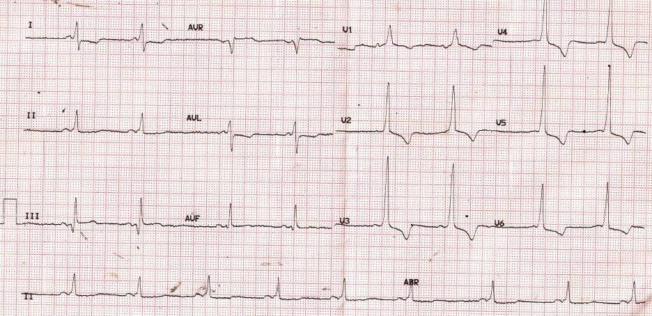
At one look it may look like left ventricular hypertrophy with strain pattern as there are tall R waves in lateral leads with ST segment depression and T wave inversion. One might think of old inferior wall infarction in addition as there is a prominent ‘Q’ wave in lead III and a smaller one in aVF. Some may even consider right ventricular hypertrophy in view of dominant R waves in V1, V2.
But if you go by the standard sequence of ECG interpretation – P, PR interval, QRS, ST segment and T wave, it becomes obvious that PR interval is short. In many leads, the slurred initial part of QRS (delta wave) is also evident (check out lead I, II and V1). Short PR interval is difficult to discern in leads III and aVF as initial part of the QRS is almost isoelectric. Taking all these findings together, the diagnosis is now obvious: Wolff-Parkinson-White Syndrome.
Wolff-Parkinson-White Syndrome is characterised by short PR interval, delta wave and secondary ST-T changes. It can predispose to tachyarrhythmias, most common being atrioventricular reentrant tachycardia (PSVT). Wolff-Parkinson-White Syndrome is due to accessory pathways which bypass the AV node and connect the atria to the ventricles with a faster bypass tract.
Related Posts


About The Author
Johnson Francis
Former Professor of Cardiology, Calicut Govt. Medical Kozhikode, Kerala, India. Editor-in-Chief, BMH Medical Journal