ECG Quiz 55 – Discussion – 3:1 AV Block
ECG Quiz 55 – Discussion – 3:1 AV Block
ECG Quiz 55 – 3:1 AV Block
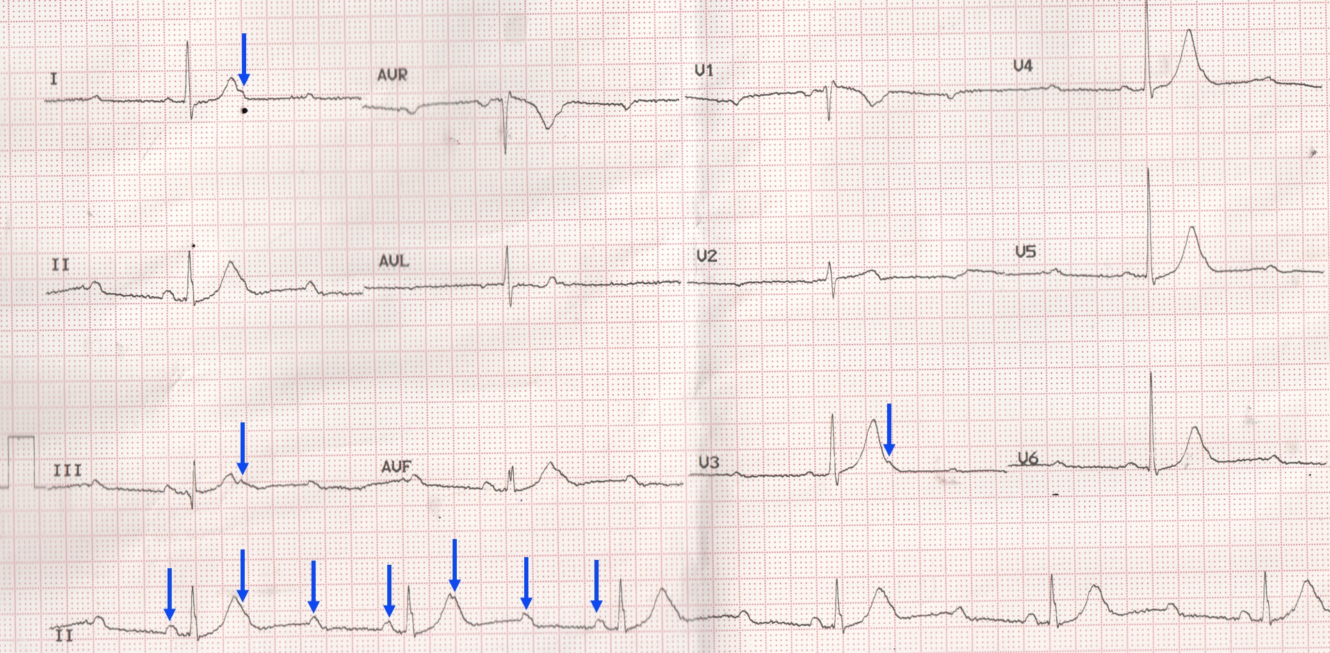
There are three P waves for every QRS complex. Two P waves between the QRS complexes are evident while one is ‘hidden’ over the T wave. Hence this is a 3:1 AV block. At an initial look, it may be called 2:1 AV block because there are only two obvious P waves between any pair of QRS complexes. But the clue is the PP interval, which is only one third of the RR interval. Then one has to look meticulously for the ‘hidden P waves’ over the T waves. The hidden P waves may not be very evident in some leads while it may be detected easily in other leads.
The QRS is not significantly widened, though there is rSr’ pattern in V1. Hence the level of the block is likely to be supraHisian. 3:1 AV block comes under advanced AV block (or high grade AV block) where two or more consecutive P waves are non conducted. Though it comes under second degree AV block in the simple classification, actually it may be considered midway between the usual second degree AV block and complete heart block.
A study by Strasberg B et al evaluated the natural history of 56 patients with chronic second degree AV block. 19 patients had no organic heart disease. Of these 7 were trained athletes. All had type I AV block and 5 had episodes of 2:1 AV block [1].
One patient without organic heart disease needed permanent pacemaker for brady arrhythmic symptoms. Two patients had non sudden death.
Among those with organic heart disease, pacemaker was implanted in ten. 16 patients died, 3 suddenly, 7 with congestive heart failure, 2 of acute myocardial infarction and 4 of non-cardiac causes.
Overall, the prognosis is poor in second degree AV block if there is associated organic heart disease. Prognosis is good in those without structural heart disease, especially in those with type I second degree AV block without bundle branch block. Type II second degree AV block with associated bundle branch block has poorer prognosis.
Reference
- B Strasberg, F Amat-Y-Leon, R C Dhingra, E Palileo, S Swiryn, R Bauernfeind, C Wyndham, K M Rosen. Natural history of chronic second-degree atrioventricular nodal block. Circulation. 1981 May;63(5):1043-9.
Related Posts


About The Author
Johnson Francis
Former Professor of Cardiology, Calicut Govt. Medical Kozhikode, Kerala, India. Editor-in-Chief, BMH Medical Journal