Old inferior wall myocardial infarction
ECG Quiz 9
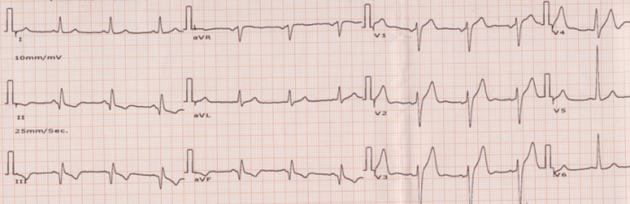
ECG shows old inferior wall infarction as evidenced by pathological Q waves in inferior leads II, III and aVF. Q waves are considered pathological if they are at least 40 msec wide and when the amplitude is more than 25 % of the ensuing R wave, former being more specific. Pathological Q waves can be seen in myocardial infarction and hypertrophic cardiomyopathy. Narrow deep Q waves in the lateral leads are seen in left ventricular volume overload. The Q waves in inferior wall infarction may disappear as time passes so that the ECG may become near normal months or years after an inferior wall infarction. This is more likely when the size of the infarct is smaller. It is less likely to occur in anterior wall infarction, though not unknown. The T wave inversions may also disappear as time passes.
Related Posts


About The Author
Johnson Francis
Former Professor of Cardiology, Calicut Govt. Medical Kozhikode, Kerala, India. Editor-in-Chief, BMH Medical Journal