ECG Quiz – Discussion
What are the findings? Can they predispose to any arrhythmia?
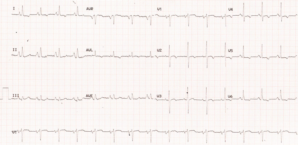
ECG shows sinus tachycardia with P pulmonale (tall peaked P waves in lead II, III and aVF) and shallow lateral T inversion. P pulmonale indicates right atrial enlargement and an enlarged atrium can predispose to atrial fibrillation (see below), which may be initially paroxysmal, but later on becomes persistent, if the atrial enlargement is not reversible. The negative P waves in V1 in this case are not due to left atrial enlargement, but due to pseudo left atrial enlargement pattern, which is often seen with right atrial enlargement. It is differentiated from true left atrial enlargement by the sharp downward slope (atrial intrinsicoid deflection). In true left atrial enlargement the downward slope is shallow the the negative P wave has more of a rounded contour.
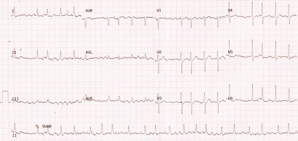
Coarse atrial fibrillation indicating underlying atrial enlargement, with a fast ventricular rate. This time it was transient, but it is likely to recur as long the primary cause of atrial enlargement is not relieved. After recurrent paroxysms, electrical and mechanical remodeling of the atrium leads to electrophysiological changes which make the atrial fibrillation permanent.
Related Posts

About The Author
Johnson Francis
Former Professor of Cardiology, Calicut Govt. Medical Kozhikode, Kerala, India. Editor-in-Chief, BMH Medical Journal