Echocardiogram in Aortic Stenosis
Echocardiogram in Aortic Stenosis
Echocardiogram is a very useful tool to assess aortic stenosis. It will identify whether the stenosis is valvar, supra valvar or subvalvar. Associated lesions like aortic regurgitation can be documented by Doppler studies. The valve morphology can be better assessed by 3-D echocardiography if available. Severity of the stenosis can be evaluated by Doppler measurement of trans valvar gradient. Alternative method is to calculate the aortic valve area by continuity equation. Indirect evidence of severity can be given by the extent of left ventricular hypertrophy and dysfunction if any.
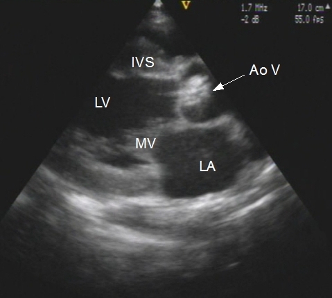
Thickened and stenosed aortic valve seen in parasternal long axis view on echocardiography. Both the mitral and aortic valves are in near closed position.
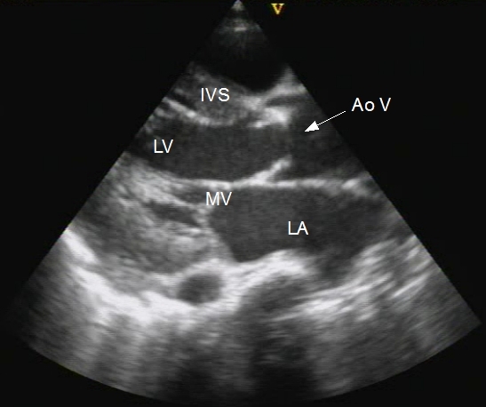
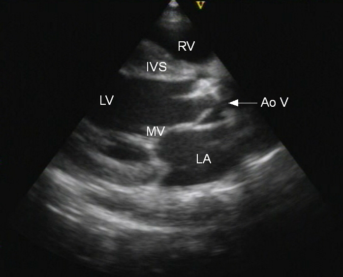
Systolic frame from an echocardiogram of aortic valve in parasternal long axis view after balloon aortic valvotomy showing restricted opening of the aortic leaflets. Upper leaflet is grossly thickened. Mitral valve is in closed position. The echocardiogram was obtained after palliative balloon aortic valvotomy prior to aortic valve replacement in a case of severe aortic stenosis with heart failure, at high risk for open heart surgery. The balloon aortic valvotomy procedure was demonstrated in a previous post. The parasternal long axis view of the open position of the aortic valve before balloon aortic valvotomy is given below.
About The Author
Johnson Francis
Former Professor of Cardiology, Calicut Govt. Medical Kozhikode, Kerala, India. Editor-in-Chief, BMH Medical Journal