Echocardiogram in Rheumatic Mitral and Aortic Regurgitation
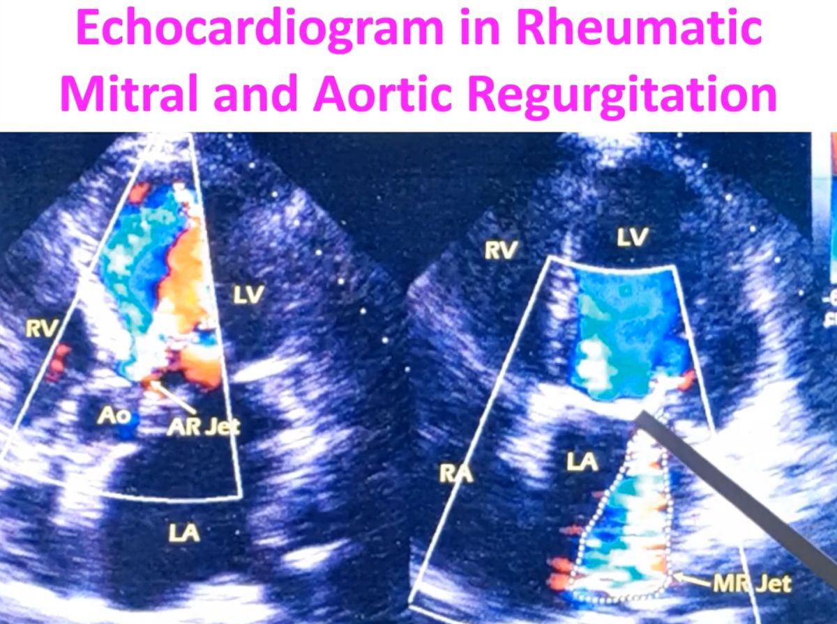
Transcript of the video: This is an apical five chamber view and this is an apical four chamber view. You can see four chambers – RV, LV, RA, LA, and the transducer location is here. And this is five chamber because, in addition you are seeing the aorta also. Right atrium has not been labelled. In this view, you can see that mitral leaflets are thickened.
 This is anterior mitral leaflet, thickened, and in the closed position of mitral valve, when there should be no flow to the left atrium, you are seeing a jet, a mosaic jet, which has been traced out. Multi-coloured jet due to high velocity and turbulence. That is what you are seeing here. This is the mitral regurgitation jet. Two things which you look at regarding the jet are, the extent to which it is extending to the left atrium, it is extending almost the whole extent, and the area of the jet, compared to the area of the left atrium. These two are useful in assessing severity. Another aspect which you can look at is the width of the jet at its origin and also this flow acceleration, before the mitral valve. That can also be looked at. As the mitral leaflets are thickened, this is likely to be due to rheumatic mitral regurgitation. And assessment of area for assessment of severity, may be erroneous if it is an eccentric jet. This is not an eccentric jet, this is almost a central jet. But if the jet is going along a wall of the left atrium, sometimes it can go along the anterior mitral leaflet into the septal side, or it can go along the posterior mitral leaflet into the lateral aspect of the left atrium. When the jet is eccentric, the assessment of area for noting the severity may not be correct. It will underestimate, if the jet is eccentric. But in a central jet, it is fairly reasonable. But colour Doppler estimation of severity is mostly semi-quantitative. You cannot say its highly quantitative estimation. Here it is the AR jet. You can see the reverse flow into the left ventricle from the aorta, with the aortic valve in closed position. This is a diastolic image, while this is a systolic image. Variance in colour because of variance in velocity and predominantly a red jet, as it is directed towards the transducer. Here it is predominantly blue because it is directed away from the transducer. This blue flow could be the flow going towards the aorta in systole. In systole, aorta is not seen here because this is only a four chamber view. This could be the flow towards the aorta. Here there is also a blue flow, but that cannot be the flow towards the aorta, as it is diastole. So most likely, this jet is going up to this and swirling around, producing a blue jet in the opposite direction. Beacuse, as the aortic valve is closed, and the regurgitant jet is coming in, and left ventricular diastolic pressures are lower than the aortic pressures. This jet cannot go, usually into the aorta. So this is probably a swirling around of the aortic regurgitation jet.

