Echocardiographic profile in ventricular septal defect – video
Echocardiographic profile in ventricular septal defect – video
Echocardiogram with narration
Echocardiogram without narration:
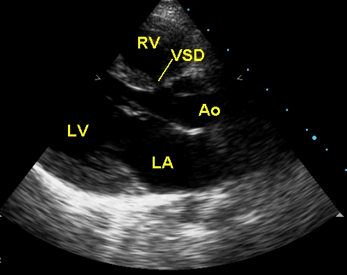
Parasternal long axis view showing aorta (Ao), left atrium (LA), left ventricle (LV) and a small perimembranous (subaortic) ventricular septal defect. Mitral valve is in the open position and the aortic valve in the closed position.
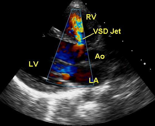
Colour sector in parasternal long axis view shows the mosaic (multi-coloured) VSD jet across the perimembranous VSD from the left ventricle to the right ventricle. It is a high velocity jet because the VSD is restrictive. The neck of the jet almost corresponds to the size of the VSD. VSD jet is seen in a systolic frame.
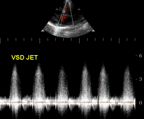
VSD jet can be picked up in parasternal long axis or short axis view, guided by color Doppler. It may also be picked up from the apical four chamber view, but the alignment may not be good. Pulsed Doppler cannot measure the jet velocity as it is much higher than the Nyquist limit of the pulsed Doppler system. Hence continuous wave Doppler is used for interrogation of the VSD jet. The interventricular gradient is calculated using the Bernoulli equation. A high interventricular gradient indicates that the VSD is restrictive. A low gradient indicates unrestrictive VSD and pulmonary hypertension.
Related Posts

About The Author
Johnson Francis
Former Professor of Cardiology, Calicut Govt. Medical Kozhikode, Kerala, India. Editor-in-Chief, BMH Medical Journal