EP catheters in LAO view
EP catheters in LAO view
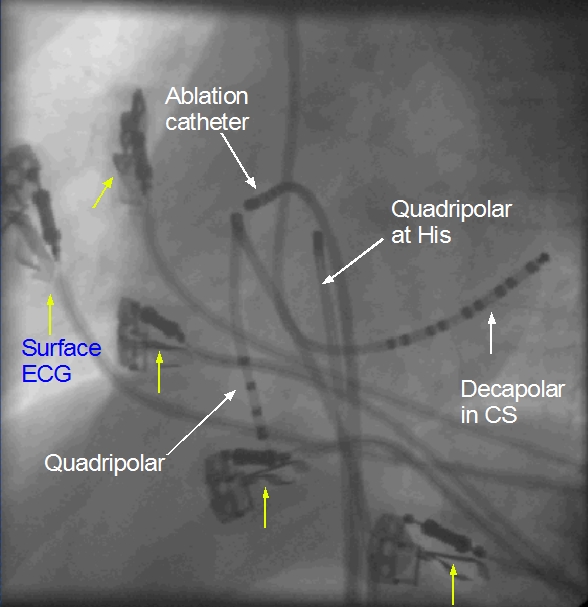
Electrophysiology (EP) catheters are shown in left anterior oblique view. The surface ECG electrodes are marked with yellow arrows. The decapolar catheter is situated in the coronary sinus and has been introduced from above using a jugular vein puncture. The electrodes are numbered from 1 – 10 in a distal to proximal fashion so that the distal pair is 1-2 and the proximal pair is 9-10. The distal electrodes record the potential from the left atrium. One quadripolar catheter is positioned in the region of the bundle of His another in the right ventricle. All electrode pairs are connected to a junction box and through it to the EP recorder. Recordings from all electrode pairs are displayed on the EP recorder. Right ventricular catheter is also used for the pacing protocols while inducing tachycardia. His bundle electrode pairs are named His proximal (Hisp) and His distal (Hisd). The pattern of activation during sinus rhythm is studied initially to identify the sequence of activation. Changes in the sequence with various pacing protocols are assessed. The ablation catheter is thicker and tip electrode is bigger with more surface area for the delivery of the radiofrequency energy during radiofrequency catheter ablation.
Related Posts

About The Author
Johnson Francis
Former Professor of Cardiology, Calicut Govt. Medical Kozhikode, Kerala, India. Editor-in-Chief, BMH Medical Journal