External iliac artery occlusion seen on a digital subtraction angiogram
External iliac artery occlusion seen on a digital subtraction angiogram
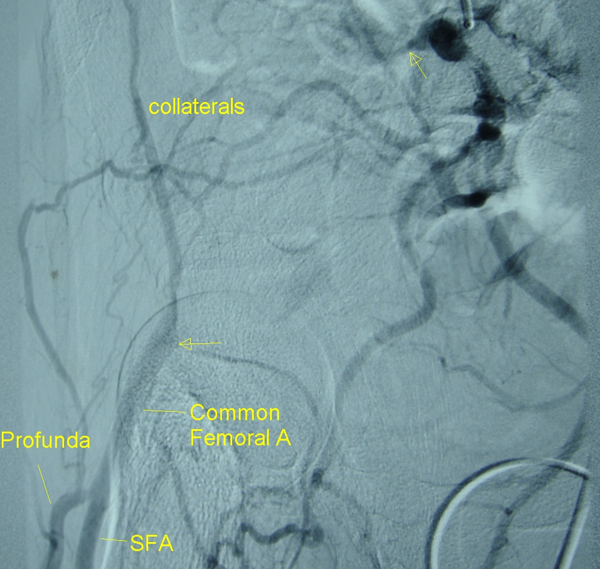
External iliac artery occlusion seen on a digital subtraction angiogram of the external iliac artery entered by the contralateral approach. Contrast is filling the internal iliac branches well and through collaterals the reformation of the common femoral artery is seen. The common femoral artery is seen as dividing into profunda femoris and superficial femoral artery lower down. The head of femur is seen in profile in the region of the common femoral artery. The upper arrow marks the probable site of external iliac artery occlusion.
Related Posts

About The Author
Johnson Francis
Former Professor of Cardiology, Calicut Govt. Medical Kozhikode, Kerala, India. Editor-in-Chief, BMH Medical Journal