Left Bundle Branch Block with Tachycardia
Left bundle branch block with tachycardia
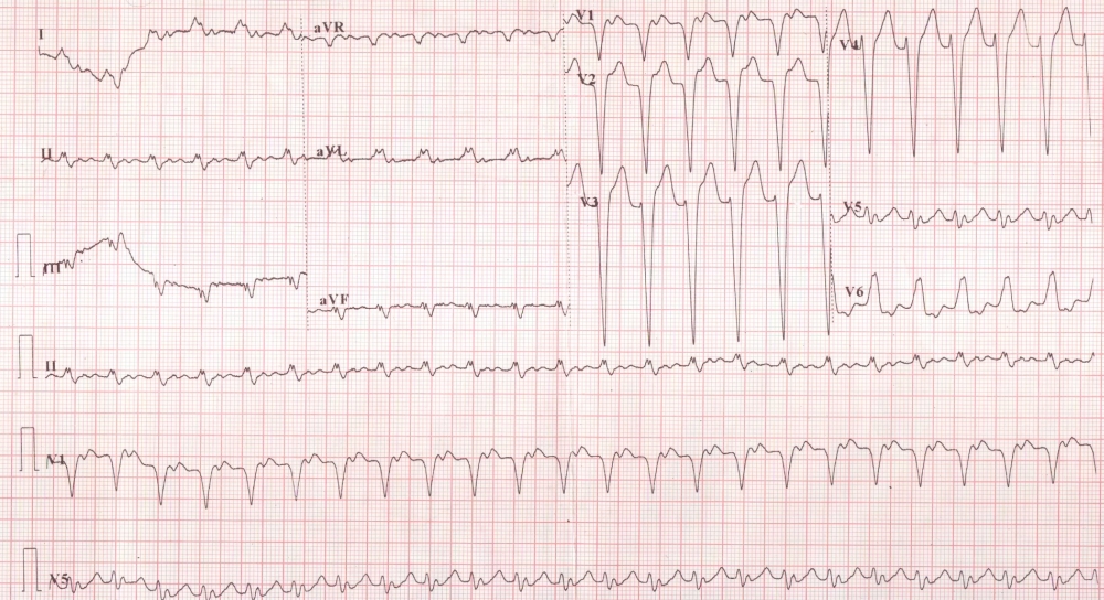
Left branch block is manifested as wide QRS with a slurred negative QRS in V1 and a slurred positive QRS in V6. The small deflection just beyond the QRS is the P wave and the next peak is the T wave. So it is evident that there is a tachycardia, possibly sinus tachycardia with a prolonged PR interval. An alternate possibility of atrial flutter with 2:1 conduction should also be thought of as the ventricular rate is around 150/minute, the classical rate for atrial flutter with 2:1 conduction. But even a close scrutiny of multiple leads do not reveal a non conducted flutter wave mid way between two proposed P waves.
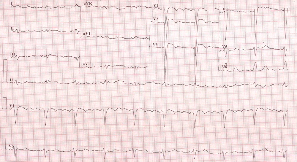
LBBB pattern is seen just as in the previous ECG. But in this tracing there are four atrial complexes for every QRS complex. The atrial rate is in the range of atrial tachycardia, nearing the flutter range. Looking back, it is possible that the upper tracing shows 2:1 conduction and the lower tracing after treatment shows 4:1 conduction because of enhancement of AV block due to the effect of the pharmacological agent which was administered in between.
About The Author
Johnson Francis
Former Professor of Cardiology, Calicut Govt. Medical Kozhikode, Kerala, India. Editor-in-Chief, BMH Medical Journal