LVH on echocardiogram
LVH on echocardiogram
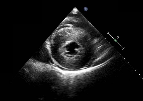
Parasternal short axis view at the level of the papillary muscles showing severe concentric left ventricular hypertrophy. Serial short axis views have to be obtained and maximal thickness documented, when assessing hypertrophic cardiomyopathy for risk profile.
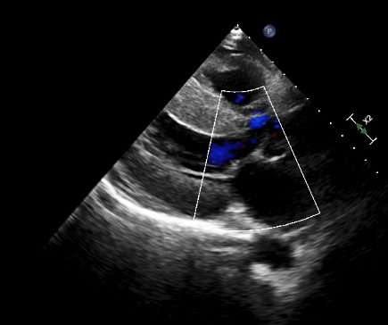
Parasternal long axis view showing concentric left ventricular hypertrophy.
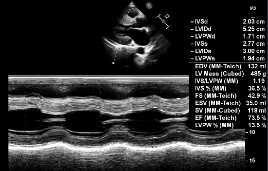
M-Mode echocardiogram showing left ventricular hypertrophy. IVSd: interventricular septum diastolic measurement. LVIDd: Left ventricular internal dimension, diastolic. LVPWd: Left ventricular posterior wall, diastolic. IVSs: Interventricular septum, systolic. LVIDs: Interventricular septum, systolic. LVPWs: Left ventricular posterior wall, systolic. EDV: End diastolic volume. FS: Fractional shortening. ESV: End systolic volume. SV: Stroke volume. EF: Ejection fraction. Here is a journal reference on detection of LVH by M-Mode echocardiogram [1].
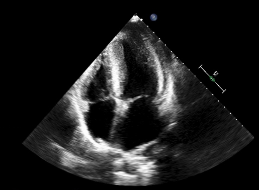
Apical four chamber view showing left ventricular hypertrophy.
Reference
- Devereux RB. Detection of left ventricular hypertrophy by M-mode echocardiography. Anatomic validation, standardization, and comparison to other methods. Hypertension. 1987 Feb;9(2 Pt 2):II19-26.
Related Posts

About The Author
Johnson Francis
Former Professor of Cardiology, Calicut Govt. Medical Kozhikode, Kerala, India. Editor-in-Chief, BMH Medical Journal