Heart Failure Awareness Week February 10-16, 2019 Heart Failure Awareness Week is celebrated by the Heart Failure Society of America (HFSA) in the week of Valentine’s day. Aim
X-ray Quiz 9 What are the findings on this post-operative chest X-ray? Some of the findings on this chest X-ray are quite obvious while some are subtle. It
ECG Quiz 47 What is the arrhythmia seen in the highlighted region of the monitor screen shot? Discussion Highlighted region shows a run of wide QRS rhythm. The
X-ray Quiz 8 Pacemaker pulse generator is seen in the right infraclavicular region. The pacemaker leads are clearly seen in this chest X-ray, either due to slight over
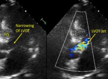
Echo quiz Discussion IVS (interventricular septum) is hypertrophied and bulges into the left ventricular outflow tract (LVOT), narrowing it. Gradient across the LVOT is increased to 26 mm
ECG Quiz 43 Discussion Rhythm strip shows sinus bradycardia with junctional escape rhythm. Junctional escape rhythm is manifested as narrow QRS complexes without preceding P waves. Second and
ECG in Ebstein’s anomaly of tricuspid valve ECG in Ebstein’s anomaly of tricuspid valve ECG in Ebstein’s anomaly of tricuspid valve showing right axis deviation of QRS, notched