PDA jet in Tetralogy of Fallot – Echocardiogram video
PDA jet in Tetralogy of Fallot – Echocardiogram video
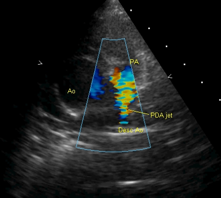
Colour Doppler imaging shows high velocity jet in the pulmonary artery arising distally, from the descending aorta, suggesting a patent ductus arteriosus. This is one of the compensatory mechanisms to improve pulmonary flow in Tetralogy of Fallot. Another mechanism is major aortopulmonary collateral arteries. Intra pulmonary collaterals can also occur in Tetralogy of Fallot. The image is in the parasternal short axis view.
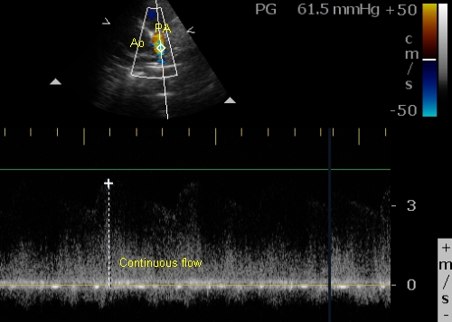
Continuous wave Doppler interrogation of the jet guided by colour flow mapping picks up the continuous flow with a peak gradient of 61.5 mm Hg. The gradient is calculated from the velocity measured by the device using the formula: V = 4 V2.
Related Posts


About The Author
Johnson Francis
Former Professor of Cardiology, Calicut Govt. Medical Kozhikode, Kerala, India. Editor-in-Chief, BMH Medical Journal