Pericardial Effusion on CXR
Pericardial Effusion on CXR
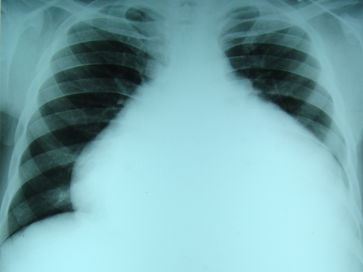
Cardiothoracic ratio is grossly increased due to the large pericardial effusion. In this case there was underlying severe right ventricular endomyocardial fibrosis as well.
Prior to the era of ubiquitous availability of echocardiography, pericardial aspiration followed by instillation of air into the pericardial cavity and taking of repeat X-ray was often done to assess the pericardial thickness. Such films could also sometimes pick up the outline of an intrapericardial mass. Computerized tomographic (CT) scans are quite useful to assess pericardial thickness. Though echocardiography is very useful for detection and quantification of pericardial effusion, it is not very accurate in assessing pericardial thickening.
Differential diagnosis of gross enlargement of cardiac size on x-ray include:
1. Large pericardial effusion: The cardiac outline is smooth in large pericardial effusion without any significant bulges in the contour. The smooth outline is due to the presence of fluid in the pericardial cavity which smoothens the outline.
2. Ebstein’s anomaly of tricuspid valve: Cardiomegaly with relative oligemia of the lung fields and reduced cardiac motion can produce a “stencilled out” appearance in Ebstein’s anomaly.
3. Multivalvular heart disease: The cardiac outline will show multiple bulges due to different chamber enlargements in multivalvular heart disease, unlike in pericardial effusion.
4. Endomyocardial fibrosis: In the classical right ventricular endomyocardial fibrosis, it is usually the grossly enlarged right atrium which enlarges the cardiac contour. There can be associated pericardial effusion as well. The prevalence of endomyocardial fibrosis has grossly declined over the past few decades.
Related Posts


About The Author
Johnson Francis
Former Professor of Cardiology, Calicut Govt. Medical Kozhikode, Kerala, India. Editor-in-Chief, BMH Medical Journal