Post BMV echocardiogram with video
Post BMV echocardiogram with video
Post BMV Echocardiogram with narration
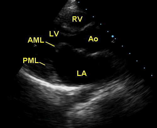
Echocardiogram in rheumatic mitral stenosis (post balloon mitral valvotomy) showing the doming of the anterior mitral leaflet (AML) and paradoxical anterior movement of the posterior mitral leaflet (PML) in diastole, indicating partial commissural fusion. Left atrium (LA) is mildly dilated. RV: right ventricle; Ao: aorta; LV: left ventricle
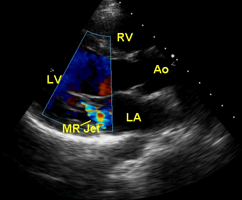
Mild mitral regurgitation is seen as a mosaic coloured jet into the left atrium in systole. The aortic valve is in the open position (systolic frame). Mitral valve is in the closed position.
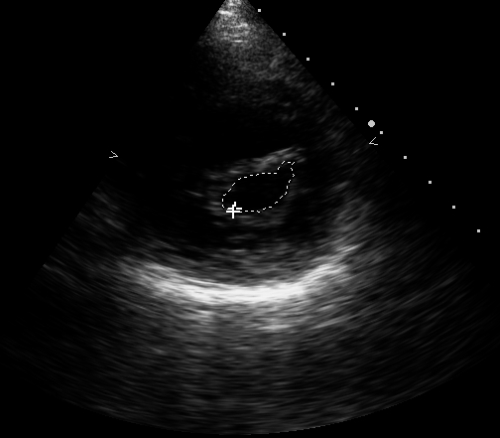
Planimetry shows good valve area after a split of the commissures with BMV (balloon mitral valvotomy).
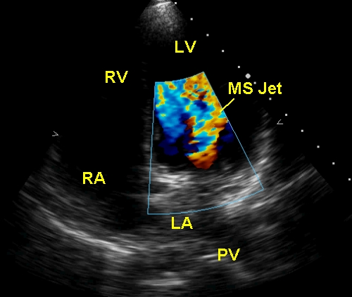
MS jet is seen as a mosaic colored jet in diastole, just beyond the mitral valve in the left ventricle. PV: pulmonary vein.
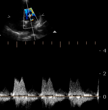
Mitral inflow Doppler tracing in mitral stenosis showing fusion of E and W waves with absent diastasis due to the persistent gradient through out diastole. But the gradients are not high in this post BMV situation. Doppler line is such that the mild mitral regurgitation has not been picked up.
About The Author
Johnson Francis
Former Professor of Cardiology, Calicut Govt. Medical Kozhikode, Kerala, India. Editor-in-Chief, BMH Medical Journal