Single chamber pacemaker and aortic prosthetic valve
Single chamber pacemaker and aortic prosthetic valve
Cardiology X-ray
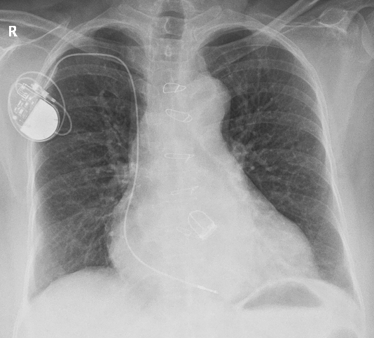
X-ray chest PA view showing a single chamber pacemaker with lead and an aortic prosthetic valve. The pulse generator of the pacemaker can be seen below the side marker (R). The pacemaker lead can be seen starting from the pacemaker header where it has been screwed in. Redundant part of the lead has been coiled within the pulse generator pocket subcutaneously as is the usual practice during pacemaker implantation. The lead can be seen coursing through the subclavian vein (just below the medial end of right clavicle, into the superior vena cava to the right atrium. The distal end of the pacemaker lead can be seen to the right of the spine near the left hemidiaphragm. The fact that the lead has crossed the midline in a PA view indicates that it has crossed the tricuspid valve which usually overlies the spine in this view. Permanent right ventricular pacing leads are often placed at the right ventricular apex which is technically easier. Several implanters now consider a position in the right ventricular outflow tract as more physiological. While pacing the right ventricular outflow tract, a screw in lead is needed to retain the position. Special lead stylets are available to negotiate the lead tip to the right ventricular outflow tract. Position in the right ventricular tract should be posteriorly in the septal aspect, avoiding the free wall of the outflow tract, to achieve the objective of early entry of signal into the conduction system. Early entry of signal into the conduction system will reduce the dyssynchrony which has been considered as a down side of right ventricular apical pacing.
Close scrutiny of the pacemaker lead tip can identify two electrodes, one at the tip and one a little proximal to that. It needs a higher resolution photograph with more penetration or ideally a fluoroscopic imaging to bring out the details of the lead tip well.
The X-ray also shows a Starr-Edwards aortic mechanical prosthetic valve and sternal suture wires. The aortic position of the valve can be identified by the location along the line joining the pulmonary bay and the right cardiophrenic angle as well as the upward direction of the cage in this case. In Starr-Edwards aortic prosthesis, the cage is seen upwards with three struts and sewing ring at the base. This contrasts with the mitral prosthesis which has four struts for the cage and the cage is directed downwards towards the left ventricular apex.
A prominent aortic knuckle and a bulge of the right atrial contour are the other findings on this chest X-ray.
About The Author
Johnson Francis
Former Professor of Cardiology, Calicut Govt. Medical Kozhikode, Kerala, India. Editor-in-Chief, BMH Medical Journal