Starr–Edwards mitral prosthetic valve on CXR
Starr–Edwards mitral prosthetic valve on CXR (X-ray Chest PA View)
Starr–Edwards mitral prosthetic valve on X-ray Chest PA view
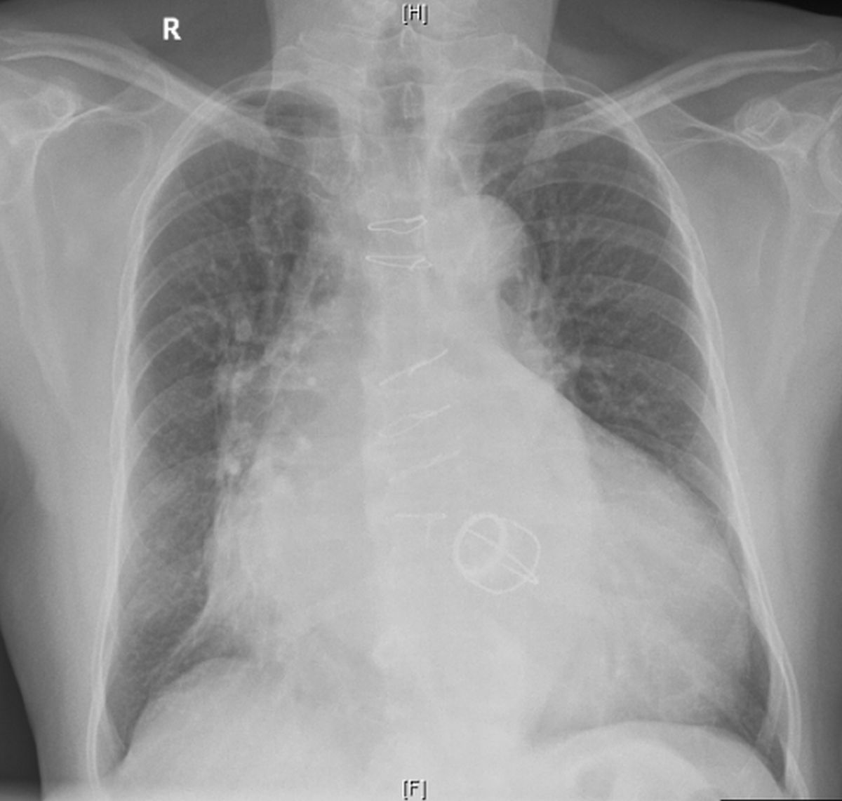
Chest X-ray PA view shows the four strut cage of Starr-Edwards prosthetic mitral valve, which is now out of production. The ring is seen towards the upper and right edge and the cage with four struts is directed downwards and left. The ball within the cage is not radiopaque enough to be seen on this chest X-ray. But it will be seen well on cine-fluoroscopy, which is used to assess the movement during cardiac cycle. Lack of good movement would indicate a stuck valve, which can lead on to acute pulmonary edema. Stuck valve could be thrombus within the valve structure, which usually occurs when the necessary anticoagulation is defective due to some reason.
Sternal wires of previous sternotomy for valve implantation are seen in the mid line. Significant cardiomegaly, biatrial enlargement and a prominent descending aortic shadow are seen. Left atrial shadow is seen as a double contour within the right cardiac border. Extension of the right cardiac border to the right indicates right atrial enlargement. Cardiac silhouette reaching almost up to the left side of the thoracic cage is indicative of left ventricular enlargement.
Starr–Edwards mitral prosthetic valve has maximum track record among mechanical prosthetic valves, functioning even after 51 years after implantation [1].
Reference
- Amrane M, Soulat G, Carpentier A, Jouan J. Starr-Edwards aortic valve: 50+ years and still going strong: a case report. Eur Heart J Case Rep. 2017 Dec 20;1(2):ytx014.
Related Posts


About The Author
Johnson Francis
Former Professor of Cardiology, Calicut Govt. Medical Kozhikode, Kerala, India. Editor-in-Chief, BMH Medical Journal