Echocardiogram in mitral stenosis
Echocardiogram in mitral stenosis
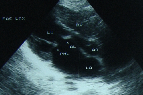
Parasternal long axis view showing dilated left atrium, thickened and paradoxically moving, that is anteriorly in diastole, posterior mitral leaflet and doming anterior mitral leaflet. The movement abnormalities of the mitral leaflets are due to commissural fusion.
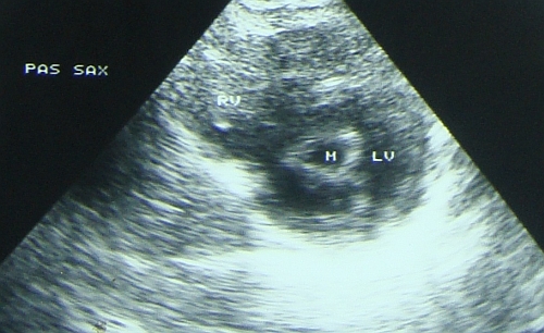
Parasternal short axis view showing the “fish mouth appearance” of the mitral orifice in the short axis view of the left ventricle. Mitral valve area can be measured in this view by planimetry using the on screen calipers.
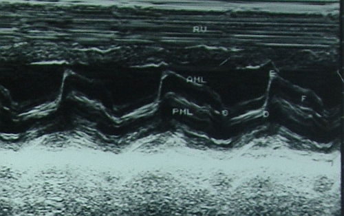
M mode echocardiogram showing the paradoxical anterior motion of posterior mitral leaflet in diastole with reduced separation of the two leaflets. Anterior mitral leaflet has certain points marked in its movement: C, D,E and F. The EF slope is reduced in mitral stenosis – it becomes almost flat in severe mitral stenosis. CD is the closed position of the mitral leaflets in systole and DE is the opening excursion of the anterior mitral leaflet.
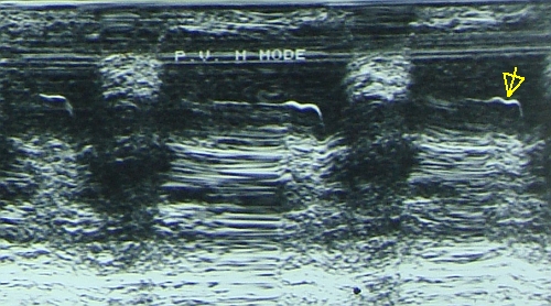
Pulmonary hypertension as evidenced by the flat EF slope of the pulmonary valve M mode echocardiogram and the shallow a wave. A mid systolic notch may also appear in severe pulmonary hypertension. When Doppler echo was not available, M mode of the pulmonary valve was an important tool to assess pulmonary hypertension. Now with Doppler echo, the right ventricular systolic pressure and indirectly the pulmonary artery systolic pressure in the absence of pulmonary stenosis, can be estimated from the velocity of the tricuspid regurgitation jet.
Related Posts


About The Author
Johnson Francis
Former Professor of Cardiology, Calicut Govt. Medical Kozhikode, Kerala, India. Editor-in-Chief, BMH Medical Journal