WPW Syndrome – the great mimicker
WPW Syndrome – the great mimicker
WPW Syndrome the great mimicker
Click here for a larger image
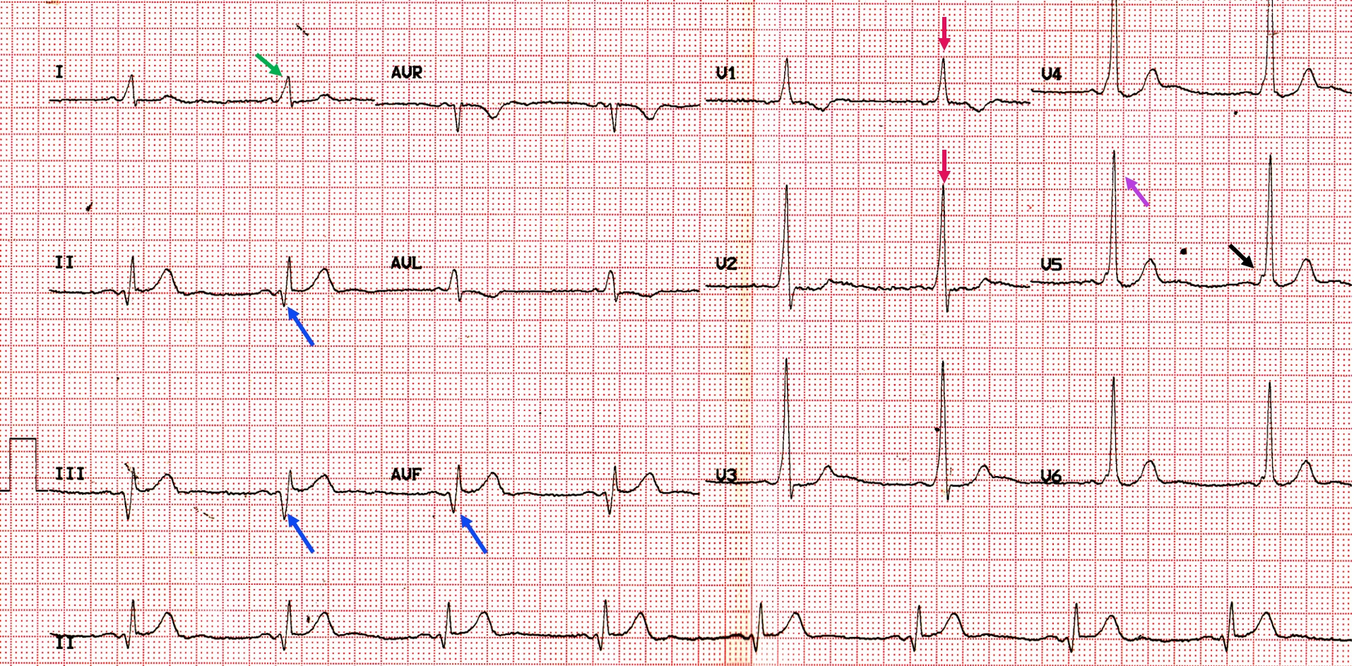
This ECG shows sinus bradycardia with short PR interval and delta waves (black arrow in V5) indicating Wolff-Parkinson-White (WPW) syndrome. WPW syndrome is a great mimicker as shown in this ECG:
- Myocardial infarction: Negative delta in inferior leads (blue arrows) may be mistaken for pathological Q waves of old myocardial infarction (pseudoinfarction pattern).
- Right ventricular hypertrophy: Tall R waves in V1, V2 (red arrows) may be mistaken for right ventricular hypertrophy.
- Left ventricular hypertrophy: Tall R waves in V5 (violet arrow) may be taken as evidence of left ventricular hypertrophy.
- Left bundle branch block: Slurred R waves in lead I (green arrow) may be considered as left bundle branch block. In fact the build in computer algorithm of the ECG machine diagnosed this ECG as ‘complete left bundle branch block’.
All these mistakes can be avoided by systematic evaluation of the ECG which will pick up the short PR interval and delta waves, which makes the actual diagnosis evident. It is the pre-excitation which causes the delta wave. Negative delta waves can be mistaken for
WPW syndrome is due to atrioventricular accessory pathways which bypass the delay of the atrioventricular node and cause pre-excitation of the ventricle. It is the pre-excitation which causes the delta wave. Negative delta waves may be mistaken for Q waves as in this case. Positive delta waves can contribute to prominent R waves as seen in V1 and mimic right ventricular hypertrophy. Widening of the QRS in lateral leads with a positive delta wave mimics left bundle branch block.
Related Posts


About The Author
Johnson Francis
Former Professor of Cardiology, Calicut Govt. Medical Kozhikode, Kerala, India. Editor-in-Chief, BMH Medical Journal