Anatomy of right atrium
Anatomy of right atrium
Right atrium is the right upper chamber of the heart. It receives de-oxygenated blood from the great veins. Superior vena cava joins it from the upper part, bringing blood from the head and neck and upper limbs. Inferior vena cava joins it from the lower part, bringing blood from abdominal organs and lower limbs. Coronary sinus bringing blood from the coronary veins joins medially near the tricuspid valve. In addition there are Thebesian veins draining the heart directly. Eustachian valve is at the orifice of the inferior vena cava and Thebesian valve at the orifice of the coronary sinus. These are vestigial structures and not complete venous valves.
Right atrium has an ellipsoid shape and the right auricle or right atrial appendage arises from it anteriorly. Interatrial septum separates the right atrium from the left atrium. Fossa ovalis in the interatrial septum is the site of fossa ovalis type of secundum atrial septal defects. A patent foramen ovale may be found in some individuals. It is a valvular opening which can shunt from right to left transiently when the right atrial pressure rises with Valsalva maneuver. Continuous shunting can occur when the foramen ovale is stretched open by enlargement of right or left atrium. In fetal life, the Eustachian valve directs inferior vena caval blood with higher oxygen saturation to the left atrium across the foramen ovale. Foramen ovale closes soon after birth when the left atrial pressure rises with functioning of the lungs and consequent increase in pulmonary venous return.
Crista terminalis is a ridge which divides the right atrium to an anterior part which has pectinate muscles and a posterior smooth part. Right atrial appendage and the region of right atrium anterior to the crista terminals is derived from the primitive atrium. Atrial pacing leads are usually placed in the right atrial appendage. Posterior part is derived from the embryonic sinus venosus and has the openings of the great veins. Sinus node is situated subepicardially, in the upper part near the opening of the superior vena cava. Atrioventricular node is in the lower part, near the opening of the coronary sinus. It lies at the apex of the triangle of Koch [1], bounded by tendon of Todaro, septal leaflet of tricuspid valve and coronary sinus orifice. Atrioventricular node is a subendocardial structure located in the lower paraseptal region.
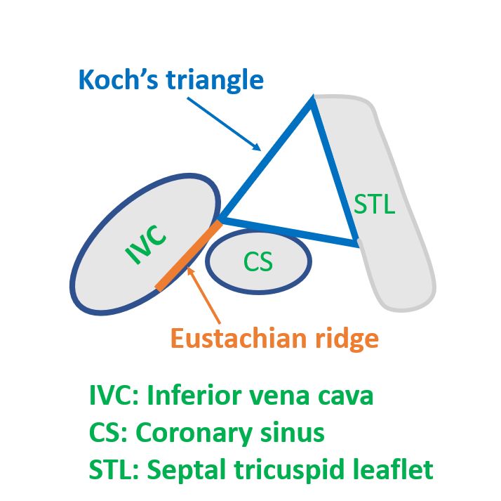
Tendon of Todaro is a collagenous subendocardial band in the right atrium. It constitutes a part of the fibrous skeleton of the heart. Tendon of Todaro runs from the central fibrous body to the Eustachian ridge. Macroscopically, tendon of Todaro is visible only in the fetal and infant heart as a white structure. It involutes later and is visible only microscopically in the adult heart. One study found tendon of Todaro visible macroscopically in only 10.8% of the hearts, that too only in the younger specimens [2]. So, in that study, they used a line joining the apex of the Koch’s triangle to the point where the basal edge of the triangle touches the Eustachian ridge. Apex of the triangle was found by transillumination of the membranous septum from the left ventricular side. Eustachian ridge separates the orifice of the inferior vena cava from the coronary sinus [3].
Blood supply to the right atrium is primarily form the right coronary artery. Conus artery is the first branch of the right coronary artery. Second branch is usually the sinus node artery. Acute marginal branches of the right coronary artery also supplies the right atrium. Right atrium forms the right border of the heart in chest X-ray PA view, located just to the right of the spine.
Right atrial enlargement is noted on chest X-ray as rightward shift of the right border of the heart. It is called as a right sided mogul. Chiari network is a vestigial structure sometimes seen in right atrium on echocardiogram. Prominent Eustachian valve may also be noted on echocardiogram sometimes. Triangle of Koch is important during catheter ablation of atrioventricular nodal re-entrant tachycardia. Ablation within the triangle of Koch has a risk of injury to the AV node and complete AV block [2].
References
- Shanubhogue S, Mohamed T, Shankar N. Morphometry of the triangle of Koch and position of the coronary sinus opening in cadaveric fetal hearts. Indian Heart J. 2017 Jan-Feb;69(1):125-128.
- Klimek-Piotrowska W, Holda MK, Koziej M, Salapa K, Piatek K, Holda J. Geometry of Koch’s triangle. Europace. 2017 Mar 1;19(3):452-457.
- McKay T, Thomas L. Prominent crista terminalis and Eustachian ridge in the right atrium: Two dimensional (2D) and three dimensional (3D) imaging. Eur J Echocardiogr. 2007 Aug;8(4):288-91.
Related Posts


About The Author
Johnson Francis
Former Professor of Cardiology, Calicut Govt. Medical Kozhikode, Kerala, India. Editor-in-Chief, BMH Medical Journal