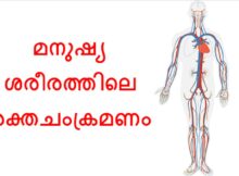
മനുഷ്യശരീരത്തിലെ രക്തചംക്രമണം ഉപാപചയ പ്രവർത്തനത്തിന് ഓക്സിജനും പോഷകങ്ങളും കോശങ്ങളിലേക്ക് കൊണ്ടുപോകുന്നതിന് അത്യന്താപേക്ഷിതമാണ്. രക്തം കാർബൺ ഡൈഓക്സൈഡും മെറ്റബോളിസത്തിന്റെ മാലിന്യ ഉൽപന്നങ്ങളും വിസർജ്ജന അവയവങ്ങളിലേക്ക് തിരികെ കൊണ്ടുപോകുന്നു. മസ്തിഷ്കം പോലുള്ള ചില അവയവങ്ങൾ ഓക്സിജൻ അടങ്ങിയ രക്തത്തെ വളരെയധികം ആശ്രയിക്കുന്നു,
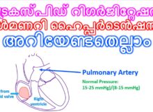
ഹൃദയത്തിന്റെയും ശ്വാസകോശത്തിന്റെയും ആരോഗ്യവുമായി ബന്ധപ്പെട്ട് നിൽക്കുന്ന ഗൗരവകരമായ രണ്ട് അവസ്ഥകളാണ് ട്രൈകസ്പിഡ് റിഗർജിറ്റേഷൻ (Tricuspid Regurgitation), പൾമണറി ഹൈപ്പർടെൻഷൻ (Pulmonary Hypertension) എന്നിവ. ഇവ പലപ്പോഴും പരസ്പരബന്ധിതമായാണ് കാണപ്പെടുന്നത്. ഇവയെക്കുറിച്ച് ലളിതമായി താഴെ വിവരിക്കുന്നു: 1. ട്രൈകസ്പിഡ് റിഗർജിറ്റേഷൻ
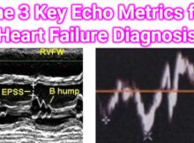
In the diagnosis of heart failure (HF), an echocardiogram is the most critical imaging tool. While a comprehensive report includes dozens of measurements, three specific metrics are considered
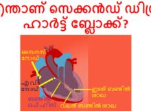
പൂർണ്ണമായ ഹാർട്ട് ബ്ലോക്ക് അഥവാ തേർഡ് ഡിഗ്രി ഹാർട്ട് ബ്ലോക്ക് നേരത്തെ കവർ ചെയ്തിട്ടുണ്ട്. സെക്കൻഡ് ഡിഗ്രി ഹാർട്ട് ബ്ലോക്കിൽ, ചില പി തരംഗങ്ങൾ മാത്രമേ വെൻട്രിക്കിളുകളിൽ എത്തുകയുള്ളൂ. ഈ ബ്ലോക്കുകൾ ഹൃദയത്തിനുള്ളിലെ വൈദ്യുതചാലകത്തിലാണെന്നും ഹൃദയ താള തകരാറുകളാണെന്നും
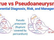
Distinguishing between a true aneurysm and a pseudoaneurysm (false aneurysm) is a critical clinical task because their underlying pathology, risk of rupture, and urgency of treatment differ significantly.
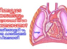
ശ്വാസതടസ്സം (Shortness of breath) അനുഭവപ്പെടുമ്പോൾ അത് ശ്വാസകോശ സംബന്ധമായ പ്രശ്നമാണോ അതോ ഹൃദയസംബന്ധമായതാണോ എന്ന് തിരിച്ചറിയുക എന്നത് വളരെ പ്രധാനമാണ്. ഹൃദയവും ശ്വാസകോശവും ഒരുമിച്ച് പ്രവർത്തിക്കുന്ന അവയവങ്ങളായതുകൊണ്ട് തന്നെ, ഒന്നിലെ തകരാർ മറ്റൊന്നിനെ ബാധിക്കാം. ഇവ തമ്മിലുള്ള
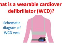
Wearable cardioverter defibrillator is a device which is worn on a vest, monitors the heart rhythm continuously and delivers a defibrillator shock in case of life threatening ventricular
ഹൃദ്രോഗത്തിന്റെ തരത്തെ ആശ്രയിച്ചിരിക്കും ലക്ഷണങ്ങൾ. ചില സമയങ്ങളിൽ രോഗലക്ഷണങ്ങളില്ലാതെ നിശബ്ദ ഹൃദ്രോഗം പോലും ഉണ്ടാകാം. ഹൃദയത്തിന്റെ ജനന വൈകല്യങ്ങളിൽ പലതും ദീർഘകാലത്തേക്ക് ലക്ഷണമില്ലാതെ തുടരുന്നു.ചിലരിൽ മറ്റൊരു രോഗാവസ്ഥയിലോ വലിയ സമ്മർദപൂരിതമായ സാഹചര്യത്തിലൊ രോഗലക്ഷണങ്ങൾ പ്രത്യേക്ഷപ്പെടുന്നു. ചിലപ്പോൾ നിശബ്ദമായ ഹൃദ്രോഗത്തിന്റെ
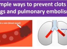
Pulmonary embolism is obstruction of blood vessels of the lungs by clots carried by blood circulation, usually from the legs. It is a serious life threatening condition and
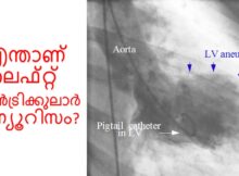
ലെഫ്റ്റ് വെൻട്രിക്കുലാർ അന്യൂറിസം ഹൃദയത്തിന്റെ ബാക്കി ഭാഗങ്ങൾ ചുരുങ്ങുമ്പോൾ താഴത്തെ ഇടത് അറയുടെ ഒരു ഭാഗം പുറത്തേക്ക് തള്ളുന്നതാണ്. ഇത് ഹൃദയാഘാതത്തിന്റെ ഒരു കോംപ്ലിക്കേഷൻ ആണ്. വീർക്കുന്ന ഭാഗത്തേക്കുള്ള രക്തധമനികൾ പൂർണ്ണമായി അടഞ്ഞിരിക്കുകയും കൊളാറ്ററൽ രക്ത വിതരണം വളരെ