Cardiac tumours
Cardiac tumours
ഹൃദയത്തിന്റെ ട്യൂമറുകൾ – മിക്കപ്പോഴും ക്യാൻസറല്ല
दिल का ट्यूमर – अक्सर कैंसर नहीं होता
Cardiac tumours are most often secondaries from malignancies of breast, lung or malignant melanoma. Primary tumours of the heart are most often benign, of which about half are myxomas. Malignant primary tumours of the heart contribute to about a quarter of the primary cardiac tumours. The commonest primary malignant tumour of the heart would be a sarcoma [1]. Cardiac tumours may present with cardiovascular or constitutional symptoms. Sometimes they are incidentally detected on echocardiography or other imaging modalities.
Computed tomography and magnetic resonance imaging can give additional information during the evaluation of cardiac tumours. CT has better spatial resolution than MRI and is useful for staging and treatment planning, especially for surgical resection of the tumour. CT also provides information on vascularity and calcification. MRI provides information about vascularity and has better tissue characterization and has no radiation risk [1].
A review mentioned that angiosarcoma is the most common malignant cardiac tumour contributing to 30% while rhabdomyosarcoma contributed 20% [2]. They also mentioned that about 10% of all tumour patients develop cardiac metastases, but they seldom manifest clinically.
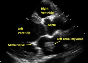
About three fourths of myxomas arise in the left atrium, while about a fifth arise in the right atrium. Ventricular myxomas are still rarer. Myxomas are usually pediculated tumours so that a left atrial myxoma can prolapse into the left ventricle in diastole as demonstrated in the YouTube video here. Majority of myxomas are sporadic and do not recur after a complete resection.
Familial myxomas can occur as a part of Carney complex. They may be associated with lentigines, cutaneous myxomas and endocrine hyperfunction. It is linked with mutation of tumour suppressor gene PRKAR1A (protein kinase A regulatory subunit-1-alpha gene) on chromosome 17q22–24. The LAMB syndrome comprises of mucocutaneous lentigines, atrial myxomas, mucocutaneous myxomas and blue nevi [3]. Components of NAME syndrome are nevi, atrial myxomas, myxoid fibromas and ephelides [4].
Papillary fibroelastoma is a rare benign tumour contributing about 5% and involves the valvular endocardium. Though they have minimal hemodynamic effects, they have a propensity for embolization. They occur in the older age group with mean age of patients being about 60 years [5].
Commonest primary cardiac tumour in children is rhabdomyoma, which is rare in adults. They occur often in the myocardium of left ventricle or interventricular septum. Rhabdomyomas may be associated with tuberous sclerosis or congenital heart disease and are often multiple. Spontaneous regression is common and can occur in about half of them. In a report of pediatric cardiac tumours over thirty years, 20 with rhabdomyomas were managed conservatively [6].
Sarcomas constitute about three fourths of primary malignant tumours of the heart. They can occur in any chamber but are frequently found in the right atrium. Life expectancy is limited to a few months without treatment. Different types are angiosarcoma, rhabdomyosarcoma, fibrosarcoma and leiomyosarcoma [2].
As mentioned initially, secondary cardiac tumours are much more common than primary cardiac tumours. Metastatic spread could be hematogenous or lymphogenous. Symptoms are often dominated by those of the primary tumour. Cardiac metastases are usually small and multiple [2].
References
- Bruce CJ. Cardiac tumours: diagnosis and management. Heart. 2011 Jan;97(2):151-60. doi: 10.1136/hrt.2009.186320. PMID: 21163893.
- Hoffmeier A, Sindermann JR, Scheld HH, Martens S. Cardiac tumors–diagnosis and surgical treatment. Dtsch Arztebl Int. 2014 Mar 21;111(12):205-11. doi: 10.3238/arztebl.2014.0205. PMID: 24717305; PMCID: PMC3983698.
- Rhodes AR, Silverman RA, Harrist TJ, Perez-Atayde AR. Mucocutaneous lentigines, cardiomucocutaneous myxomas, and multiple blue nevi: the “LAMB” syndrome. J Am Acad Dermatol. 1984 Jan;10(1):72-82. doi: 10.1016/s0190-9622(84)80047-x. PMID: 6693605.
- Atherton DJ, Pitcher DW, Wells RS, MacDonald DM. A syndrome of various cutaneous pigmented lesions, myxoid neurofibromata and atrial myxoma: the NAME syndrome. Br J Dermatol. 1980 Oct;103(4):421-9. doi: 10.1111/j.1365-2133.1980.tb07266.x. PMID: 7437308.
- Mariscalco G, Bruno VD, Borsani P, Dominici C, Sala A. Papillary fibroelastoma: insight to a primary cardiac valve tumor. J Card Surg. 2010 Mar;25(2):198-205. doi: 10.1111/j.1540-8191.2009.00993.x. Epub 2010 Feb 9. PMID: 20149002.
- Günther T, Schreiber C, Noebauer C, Eicken A, Lange R. Treatment strategies for pediatric patients with primary cardiac and pericardial tumors: a 30-year review. Pediatr Cardiol. 2008 Nov;29(6):1071-6. doi: 10.1007/s00246-008-9256-6. Epub 2008 Jul 4. PMID: 18600370.
Related Posts


About The Author
Johnson Francis
Former Professor of Cardiology, Calicut Govt. Medical Kozhikode, Kerala, India. Editor-in-Chief, BMH Medical Journal