ST prolongation in hypocalcemia ST prolongation in hypocalcemia: ST prolongation as a cause of QT interval prolongation is less common than other mechanisms of QT prolongation. In most
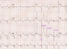
VVI pacing with ectopic beat and fusion beat VVI pacing with ectopic beat and fusion beat: Basic rhythm in this ECG is a paced rhythm, with pacing spikes
RBBB, LAHB, Junctional rhythm RBBB with LAHB & Junctional rhythm: ECG shows a slow regular rhythm with a ventricular rate of around 43/min. Though there are some baseline
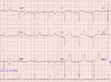
Old inferior wall infarction and lateral ST depression ECG shows sinus rhythm at around 75/min, with pathological Q waves in inferior leads, indicating old inferior wall myocardial infarction.
Monomorphic ventricular premature complex (VPC) Basic rhythm in the ECG is sinus rhythm at around 60/min. There are two wide QRS complexes seen in lead II rhythm strip.
Old inferior wall infarction and lateral ST depression ECG shows sinus rhythm at around 75/min, with pathological Q waves in inferior leads, indicating old inferior wall myocardial infarction.
Evolved anterior wall myocardial infarction ECG shows sinus rhythm at a rate of around 100/min, with QS complexes in anterior leads along with a coved ST segment elevation
Pacing in complete heart block – ECG Pacing in complete heart block (CHB): Initial part of the ECG shows narrow QRS complexes at a rate of around 43/minute.