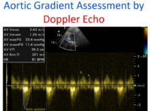
Aortic gradient assessment by Doppler echo Normal gradient across aortic valve can be measured by pulsed Doppler echocardiography as the gradients are low. In the presence of aortic
Echocardiogram in Aortic Stenosis Echocardiogram is a very useful tool to assess aortic stenosis. It will identify whether the stenosis is valvar, supra valvar or subvalvar. Associated lesions
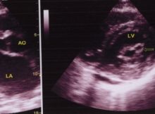
Echocardiogram in Mitral Stenosis Left panel shows the parasternal long axis view (PLAX). AO: Aorta; MVO: Mitral valve opening; LA: left atrium. Doming of the anterior mitral leaflet
Need for TEE guidance for septal puncture during BMV TEE guidance for septal puncture during BMV: Usually only fluroscopic guidance is required during trans-septal puncture to enter the
Wilkins echocardiographic score for mitral stenosis Wilkins echocardiographic score for mitral stenosis is useful for deciding whether the valve is suitable for balloon mitral valvotomy (BMV), also known
Peak vs mean trans-mitral gradient in mitral stenosis Peak trans-mitral gradient depends on the left atrial compliance and left ventricular diastolic function. Mean trans-mitral gradient reflects the severity
Quantification of tethering of mitral valve in ischemic MR Quantification of tethering of mitral valve in ischemic MR: Both two dimensional and three dimensional echocardiography are useful in
Estimation of right ventricular systolic pressure by Doppler echo is from: a) Velocity of tricuspid regurgitation jet b) Velocity of pulmonary regurgitation jet c) Area of tricuspid regurgitation