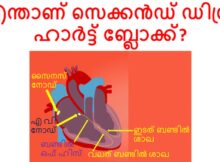
പൂർണ്ണമായ ഹാർട്ട് ബ്ലോക്ക് അഥവാ തേർഡ് ഡിഗ്രി ഹാർട്ട് ബ്ലോക്ക് നേരത്തെ കവർ ചെയ്തിട്ടുണ്ട്. സെക്കൻഡ് ഡിഗ്രി ഹാർട്ട് ബ്ലോക്കിൽ, ചില പി തരംഗങ്ങൾ മാത്രമേ വെൻട്രിക്കിളുകളിൽ എത്തുകയുള്ളൂ. ഈ ബ്ലോക്കുകൾ ഹൃദയത്തിനുള്ളിലെ വൈദ്യുതചാലകത്തിലാണെന്നും ഹൃദയ താള തകരാറുകളാണെന്നും
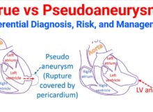
Distinguishing between a true aneurysm and a pseudoaneurysm (false aneurysm) is a critical clinical task because their underlying pathology, risk of rupture, and urgency of treatment differ significantly.
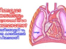
ശ്വാസതടസ്സം (Shortness of breath) അനുഭവപ്പെടുമ്പോൾ അത് ശ്വാസകോശ സംബന്ധമായ പ്രശ്നമാണോ അതോ ഹൃദയസംബന്ധമായതാണോ എന്ന് തിരിച്ചറിയുക എന്നത് വളരെ പ്രധാനമാണ്. ഹൃദയവും ശ്വാസകോശവും ഒരുമിച്ച് പ്രവർത്തിക്കുന്ന അവയവങ്ങളായതുകൊണ്ട് തന്നെ, ഒന്നിലെ തകരാർ മറ്റൊന്നിനെ ബാധിക്കാം. ഇവ തമ്മിലുള്ള
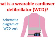
Wearable cardioverter defibrillator is a device which is worn on a vest, monitors the heart rhythm continuously and delivers a defibrillator shock in case of life threatening ventricular
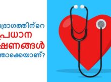
ഹൃദ്രോഗത്തിന്റെ തരത്തെ ആശ്രയിച്ചിരിക്കും ലക്ഷണങ്ങൾ. ചില സമയങ്ങളിൽ രോഗലക്ഷണങ്ങളില്ലാതെ നിശബ്ദ ഹൃദ്രോഗം പോലും ഉണ്ടാകാം. ഹൃദയത്തിന്റെ ജനന വൈകല്യങ്ങളിൽ പലതും ദീർഘകാലത്തേക്ക് ലക്ഷണമില്ലാതെ തുടരുന്നു.ചിലരിൽ മറ്റൊരു രോഗാവസ്ഥയിലോ വലിയ സമ്മർദപൂരിതമായ സാഹചര്യത്തിലൊ രോഗലക്ഷണങ്ങൾ പ്രത്യേക്ഷപ്പെടുന്നു. ചിലപ്പോൾ നിശബ്ദമായ ഹൃദ്രോഗത്തിന്റെ
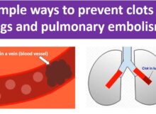
Pulmonary embolism is obstruction of blood vessels of the lungs by clots carried by blood circulation, usually from the legs. It is a serious life threatening condition and
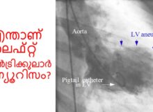
ലെഫ്റ്റ് വെൻട്രിക്കുലാർ അന്യൂറിസം ഹൃദയത്തിന്റെ ബാക്കി ഭാഗങ്ങൾ ചുരുങ്ങുമ്പോൾ താഴത്തെ ഇടത് അറയുടെ ഒരു ഭാഗം പുറത്തേക്ക് തള്ളുന്നതാണ്. ഇത് ഹൃദയാഘാതത്തിന്റെ ഒരു കോംപ്ലിക്കേഷൻ ആണ്. വീർക്കുന്ന ഭാഗത്തേക്കുള്ള രക്തധമനികൾ പൂർണ്ണമായി അടഞ്ഞിരിക്കുകയും കൊളാറ്ററൽ രക്ത വിതരണം വളരെ
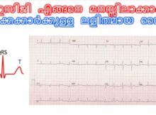
ഇസിജി (ECG/EKG) എന്നാൽ ഹൃദയത്തിന്റെ വൈദ്യുത തരംഗങ്ങളെ ഒരു പേപ്പറിലേക്ക് രേഖപ്പെടുത്തുന്ന രീതിയാണ്. ഇസിജി കൃത്യമായി വിശകലനം ചെയ്യാൻ വിദഗ്ദ്ധ പരിശീലനം ആവശ്യമാണെന്നിരുന്നാലും, ഒരു തുടക്കക്കാരൻ എന്ന നിലയിൽ അതിലെ അടിസ്ഥാന കാര്യങ്ങൾ എങ്ങനെ മനസ്സിലാക്കാം എന്ന് നോക്കാം.
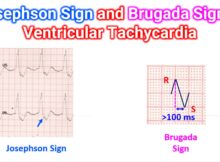
Both the Josephson Sign and the Brugada Sign are specific morphological “clues” on an ECG that help confirm a diagnosis of Ventricular Tachycardia (VT) rather than Supraventricular Tachycardia
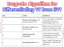
The Brugada Algorithm is for differentiation: Is this Ventricular Tachycardia or just Supraventricular Tachycardia with “aberrancy” (like a bundle branch block)? You follow these 4 steps in order.