Category: Nuclear Cardiology
Positron emission tomography
Flurpiridaz, an F-18 based agent for positron emission tomography is all set to revolutionize myocardial perfusion imaging once again.
Read More
Nuclear Cardiology
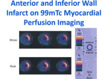
Anterior and inferior wall infarct on 99mTc myocardial perfusion imaging.
Read More
Nuclear Cardiology
Isolated reversible perfusion defects can occur in the inter ventricular septum with nuclear cardiology imaging in left bundle branch block (LBBB).
Read More
Nuclear Cardiology
Metabolic tracers for PET scan include C-11 palmitate, I-123 Beta-methyl-p-[123I]-iodophenyl-pentadecanoic acid (BMIPP) and F18 fluro-deoxy glucose (FDG).
Read More
Nuclear Cardiology
Nuclear imaging of the heart is useful in risk stratification. A normal perfusion image after adequate stress would mean a cardiac event rate of < 1%.
Read More
Positron emission tomography
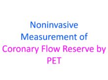
Myocardial blood flow (measured by positron emission tomography) during stress divided by resting flow was taken as the coronary flow reserve.
Read More
Positron emission tomography
Positron emission tomography (PET) can measure both the myocardial perfusion and metabolism.
Read More
Nuclear Cardiology
Rest and nitroglycerine myocardial viability scan: Rest images obtained after 8 mCi of 99mTc IV. 25 mCi hours later after S/L NTG; rest gated imaging done.
Read More
Posts navigation