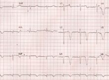
Deep T inversion First few conditions which come to our mind when we see deep T wave inversions are: Coronary artery disease Hypertrophic cardiomyopathy Post cardiac arrest state
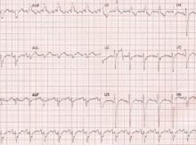
Inferolateral myocardial infarction ST segment elevation is seen in inferolateral leads (II, III, aVF, V5, V6) indicating inferolateral ST elevation myocardial infarction (STEMI). There is a discordance between
Right bundle branch block with right upper quadrant axis ECG demonstrating right bundle branch block with right upper quadrant axis: rsR’ pattern in V1, rS pattern in I,
Iodixanol is a —- radiocontrast: a) Iso-osmolar b) Low osmolar c) High osmolar d) None of the above Correct answer: a) Iso-osmolar The osmolality of iodixanol is
Contrast induced renal injury does not involve: a) Glomerulus b) Proximal convoluted tubule c) Descending loop of Henle d) Ascending loop of Henle Correct answer: a) Glomerulus
Predictors of contrast induced acute kidney injury: a) Estimated glomerular filtration rate (eGFR) b) ST elevation myocardial infarction (STEMI) c) Cardiogenic shock d) All of the above
Modified PLAX view in TGA Parasternal long axis (PLAX) view is often the first view taken during echocardiography. Usually it visualizes the left ventricle, left atrium, right ventricular
Treadmill Exercise ECG, also known as treadmill test (TMT) and stress ECG is usually done with a computerised treadmill unit which controls the motor speed of the treadmill
LV aneurysm seen on left ventriculogram and near total occlusion of the left anterior descending coronary artery (LAD), which is poorly collateralized.