Closed Loop Catheter Position in Patent Ductus Arteriosus
Closed Loop Catheter Position in Patent Ductus Arteriosus
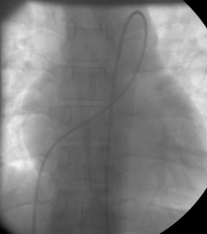
Right heart catheter is seen passing from the inferior vena cava into right ventricle through the right atrium. From the right ventricle the catheter passes into the pulmonary artery and then across the patent ductus arteriosus into the descending aorta. The position of catheter in the descending aorta is confirmed fluoroscopically by passing it below the level of diaphragm. The pressure tracing and oxygen saturation of blood withdrawn also help in differentiating it from a catheter position in the left pulmonary artery. The aortic pressure tracing will usually show a high pressure while the pulmonary artery tracing will be of lower amplitude.
But this may be different in a person with severe pulmonary hypertension. In fact in this case the pulmonary artery pressures were systemic as the PDA was quite large. Oxygen saturation will be much higher in the aorta in case of left to right shunt, but can be lower if there is significant right to left shunting across the PDA. So the only definite way to differentiate position in descending aorta from that in left pulmonary artery is by passage of the catheter below the level of left hemi-diaphragm.
Pattern of the loop is different between PDA and aortopulmonary window. Whereas it is a closed loop in PDA, it is an open loop in AP window. An open loop can also occur when the catheter passes from the right ventricle to aorta through a ventricular septal defect. PDA catheter position in lateral view is given follows.
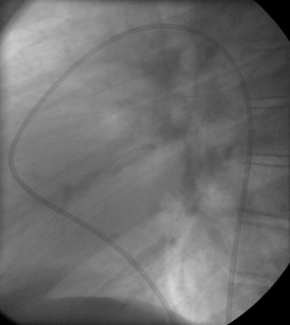
The image in lateral view shows the right heart catheter passing through patent ductus arteriosus into descending aorta. Spine is visible on right side of the screen. Right heart catheter coming through the inferior vena cava below the diaphragm is coursing anteriorly as it passes through the right atrium, right ventricle, right ventricular outflow tract and pulmonary artery. It curves posteriorly to enter ductus arteriosus and descending aorta, where it descends along the anterior border of the spine. Region of the catheter crossing tracheal air column in front of the spine superiorly is the site of patent ductus arteriosus.
Related Posts


About The Author
Johnson Francis
Former Professor of Cardiology, Calicut Govt. Medical Kozhikode, Kerala, India. Editor-in-Chief, BMH Medical Journal