Coarse trabeculae in hypertrophied right ventricle – ventriculogram
Coarse trabeculae in hypertrophied right ventricle – ventriculogram
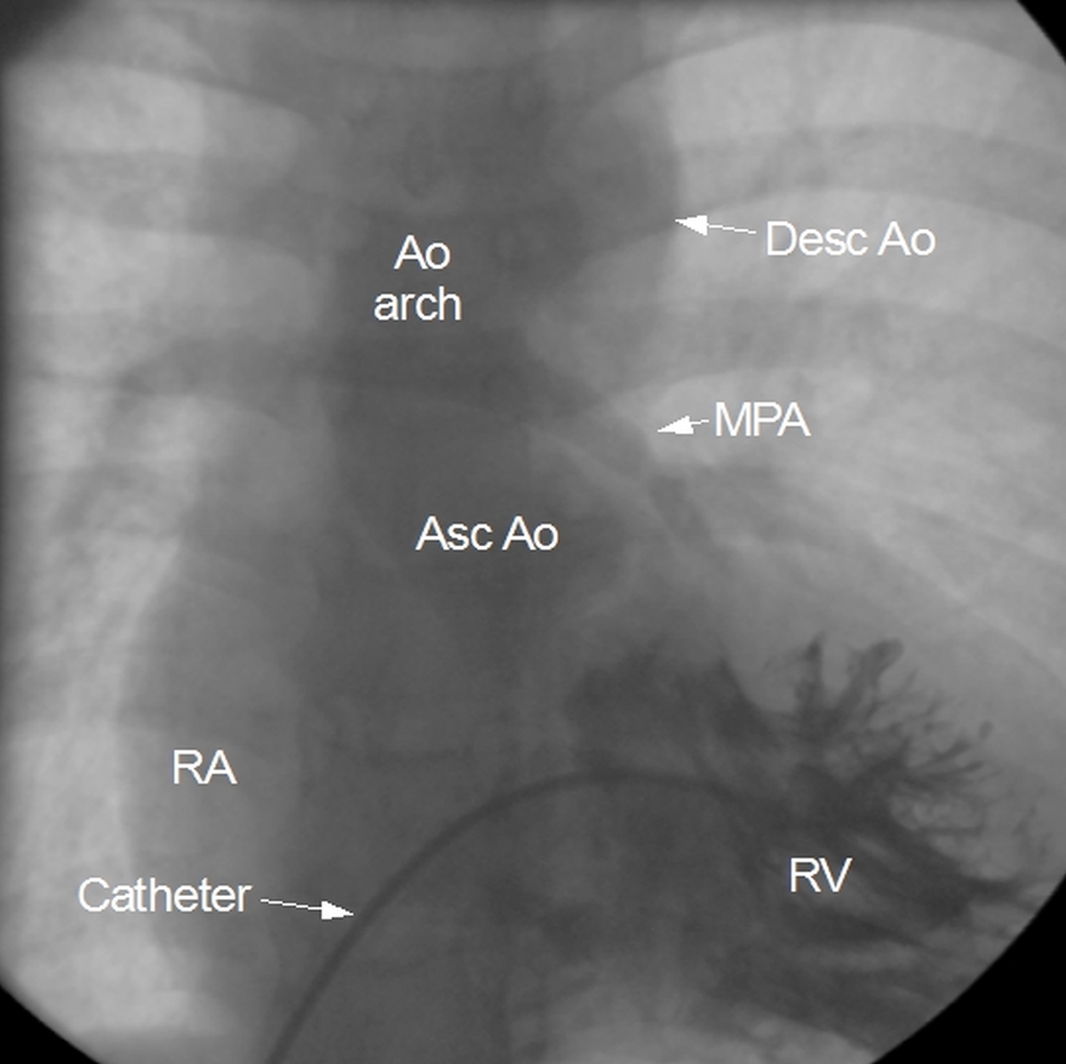
Still frame from a right ventricular angiogram showing coarse trabeculae in a hypertrophied right ventricle (negative shadows within the right ventricle). Main pulmonary artery which is faintly seen is hypoplastic. Main pulmonary artery continues as the right pulmonary artery and the left pulmonary artery is not visible (probably atretic). RA: right atrium; RV: right ventricle; Asc Ao: ascending aorta; MPA: main pulmonary artery; Ao arch: aortic arch; Desc Ao: descending aorta. The pulmonary angiogram of this case was illustrated in the previous post. A large aorta is seen filling possibly through a ventricular septal defect as in Tetralogy of Fallot. In this case the hypertrophy of right ventricle is due to outflow obstruction. Outflow obstruction can be at pulmonary arterial level (hypoplastic pulmonary arteries), pulmonary valvar level (pulmonary stenosis) or infundibular level. Sometimes the obstruction is below the infundibular level as in double chambered right ventricle (DCRV). In DCRV, a hypertrophied anomalous muscle bundle will be seen as a negative shadow much below the level of the infundibulum. Obstruction can also be at the os infundibulum, the lower part of the infundibulum.
Related Posts

About The Author
Johnson Francis
Former Professor of Cardiology, Calicut Govt. Medical Kozhikode, Kerala, India. Editor-in-Chief, BMH Medical Journal