Colour Doppler echocardiogram in anterior wall myocardial infarction – video
Colour Doppler echocardiogram in AWMI
Echocardiogram with narration:
Echocardiogram without narration:
Initial view is the parasternal long axis which shows the right ventricle above and left ventricle below, with the interventricular septum in between. The aorta and left atrium are seen to the right of image. It can be seen that the mitral and aortic leaflets are opening and closing well. There is mild thickening of the anterior mitral leaflet, but there is no doming. The contractile movements of the septum is diminished. The next view is the parasternal short axis view of the left ventricle. Contractions of the anterior wall are diminished, while the inferior, posterior and lateral walls contract well. Mitral leaflets are seen in cross section in the initial view at the level of the mitral valve while papillary muscles are seen in the distal cut. Next view is the apical four chamber view which shows all the four cardiac chambers as well as the mitral and tricuspid valves. Papillary muscle and chordae attached to the anterior mitral leaflet are also seen. The defective septal motion is evident in this view. Echo drop outs in the interatrial septum can occur in this view as the ultrasound beam is parallel to interatrial septum in this view. Pulmonary veins are seen outside the left atrium, in the right lower quadrant of the image. Apical four chamber view colour flow mapping shows the forward mitral flow in red colour. There is no mitral regurgitation. The last image is the colour flow mapping in the apical five chamber view. In the apical five chamber view, the aorta is also seen in addition to the four cardiac chambers. The opening and closing motions of the aortic valve are seen in this view. The bluish colour of the flow mapping in the aorta indicates flow away from the transducer. There is some variance in the colour due to slightly higher velocity of ejection producing some turbulence in the aorta.
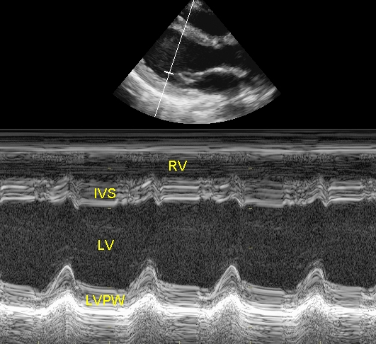
The upper image shows the 2-D echocardiogram in parasternal long axis view used for guiding the M-mode cut. The cursor is seen to cut the left ventricle at the chordal level, just beyond the tip of the mitral leaflets. The motion of the interventricular septum (IVS) is almost flat, due to the infarction. RV: right ventricle; LV: left ventricle; LVPW: left ventricular posterior wall
Related Posts


About The Author
Johnson Francis
Former Professor of Cardiology, Calicut Govt. Medical Kozhikode, Kerala, India. Editor-in-Chief, BMH Medical Journal