Diastolic dysfunction by tissue Doppler
Diastolic dysfunction by tissue Doppler
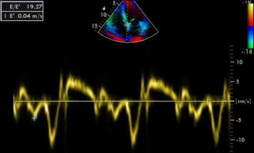
E/E’ measurement by Tissue Doppler Imaging
E/E’ measurement is used to assess diastolic function by tissue Doppler. Method may vary in technical details between machines. In low level machines, we have to measure both E from mitral flow Doppler and E’ from tissue Doppler at the septal mitral annular level and calculate the ratio manually. In other machines, as illustrated in the image above, E is measured initially and stored (not shown in the image). After that E’ is measured and the machine automatically calculates the E/E’ ratio. The high E/E’ ratio in this case indicates left ventricular diastolic dysfunction. E/E’ at lateral mitral annulus more than 10 and E/E’ at septal mitral annulus more than 15 indicates left ventricular diastolic dysfunction. E/E’ values less than 8 would indicate normal left ventricular diastolic function [1].
The yellow tracing is pulsed wave tissue Doppler imaging and ‘+’ is for the mitral E’. The next negative wave after E’ occurs during atrial contraction and is designated Aa. The positive wave after Aa is the Sa wave, representing the systolic myocardial wave recorded as the annulus descends towards the apex.
E’ velocity is also known as Ea (‘a’ for annulus) velocity or Em (‘m’ for myocardial) velocity. It reflects the early myocardial relaxation and occurs during the ascend of the mitral annulus. Septal E’ is slightly lower than lateral E’. Measurement of E’ is useful in differentiation of pseudo normalization in the mitral inflow from the normal pattern.
Reference
- Ho CY, Solomon SD. A clinician’s guide to tissue Doppler imaging. Circulation. 2006 Mar 14;113(10):e396-8.
About The Author
Johnson Francis
Former Professor of Cardiology, Calicut Govt. Medical Kozhikode, Kerala, India. Editor-in-Chief, BMH Medical Journal