Double valve replacement – echocardiographic profile with video
Double valve replacement – echocardiographic profile with video
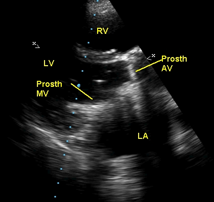
Parasternal long axis view of echocardiogram in double valve replacement. Mitral and aortic valves are prosthetic, as evident from the dense echoes due to the metallic components of the prosthetic valves. Acoustic shadowing is seen below the prosthetic aortic valve (Prosth Ao V). RV: right ventricle; LA: left atrium; LV: left ventricle. Prosth MV: prosthetic mitral valve. Left atrium is dilated, possibly as a residua of the previous lesions which made double valve replacement necessary. Aortic valve is in the closed position and the mitral valve open. The open tilting disc of the mitral valve is seen as thin echo in the lower portion of the left ventricular outflow tract, with an echodense tip.
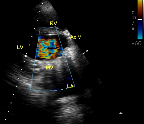
Color Doppler imaging from the parasternal long axis view shows turbulent mitral inflow through the prosthetic mitral valve. Unlike in a normal mitral valve, some turbulence is common in a normally functioning prosthetic valve. A gradient upto 5 mm Hg may be normal across a prosthetic mitral valve. The colour bar at the top right corner shows a Nyquist limit of the color flow mapping as 60 cm/s. Hence any flow with velocity above that will show aliasing. AoV: aortic valve.
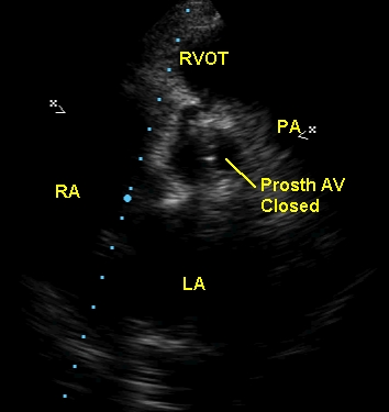
Prosthetic aortic valve in the closed position (Prosth AV Closed), seen from the parasternal short axis view. PA: pulmonary artery; RVOT: right ventricular outflow tract. In this view also the left atrium appears dilated.
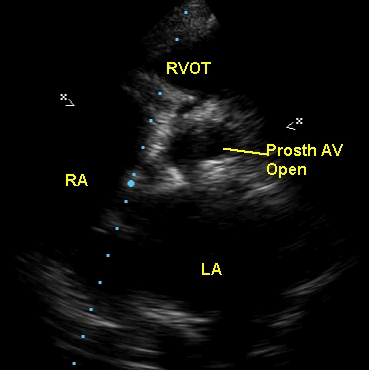
Parasternal short axis view frame showing prosthetic aortic valve in open position, with a good orifice. The shadows in the upper and lower parts of the aorta in cross section are probably the sewing ring of the prosthetic valve.
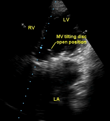
Prosthetic mitral valve with tilting disc in open position, seen on echocardiography from a modified apical four chamber view. The dense shadow below that is the ring of the prosthetic mitral valve. Movements of the disc can be assessed in this view. The angle through which the disc moves can be evaluated. Here it is about 45 degrees.
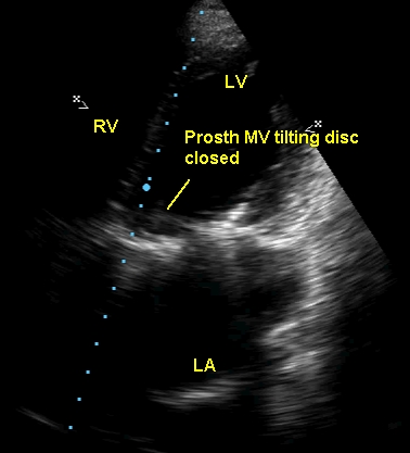
Prosthetic mitral valve with tilting disc in closed position, seen in a modified apical four chamber view.
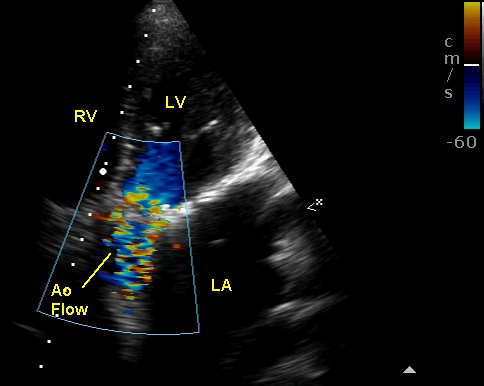
Turbulent aortic flow in prosthetic aortic valve. Some turbulence can occur even without prosthetic valve dysfunction. Basically a prosthetic valve in the aortic position is always partially obstructive, more so when a small prosthesis is placed as in aortic stenosis with a small aortic annulus. This is often called patient-prosthesis mismatch. Hence it is worthwhile documenting gradients serially so that any significant rise can alert the possibility of new obstruction developing like thrombus or pannus formation.
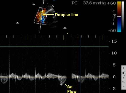
Gradient across prosthetic aortic valve measured from apical five chamber view by Doppler interrogation. The gradient is mildly elevated and 37.6 mm Hg. The gradient is calculated from the velocity using the formula: gradient = 4V2, where V is the velocity in m/s.
Related Posts


About The Author
Johnson Francis
Former Professor of Cardiology, Calicut Govt. Medical Kozhikode, Kerala, India. Editor-in-Chief, BMH Medical Journal