Can you localize the coronary lesion and explain?
ECG Quiz 1
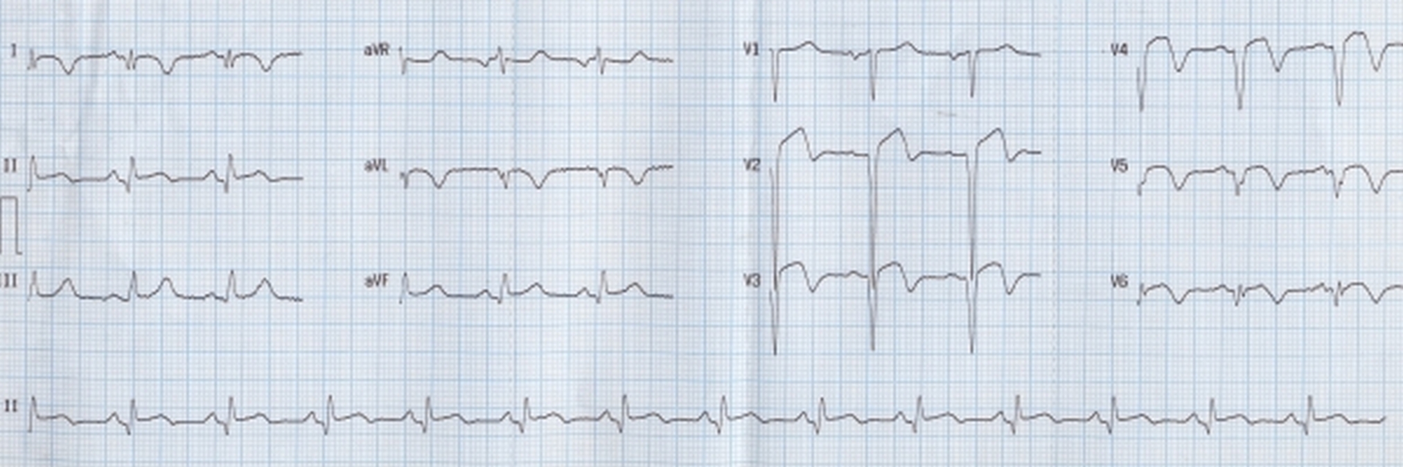
What is the diagnosis?
This ECG shows features of evolved anterior wall myocardial infarction, with additional mild ST elevation in lead II, but not in lead III.
What are the diagnostic features in this ECG?
Q waves are seen in leads I, aVL and V2 to V6. Convex upwards ST segment elevation is seen in leads V2 to V4. There is mild ST elevation in lead II. T waves are inverted in I, aVL and V2 to V6.
Can you localize the coronary lesion and explain?
In anterior wall infarction, the culprit vessel is left anterior descending coronary artery. Since V1 does not show ST elevation, the coronary obstruction is likely to be distal to first septal perforator and proximal to the major diagonal vessel. ST elevation in lead II also suggests a wrap around LAD, supplying the inferolateral region of left ventricle as well. In lesions proximal to the first septal perforator, ST elevation in inferior leads do not occur as it is canceled by anterior ST elevation.
About The Author
Johnson Francis
Former Professor of Cardiology, Calicut Govt. Medical Kozhikode, Kerala, India. Editor-in-Chief, BMH Medical Journal