Old inferior wall myocardial infarction with RBBB
Old inferior wall myocardial infarction with RBBB
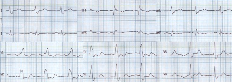
Regular sinus rhythm at about 60 per minute with a wide QRS is noted. The PR interval is 200 ms. The initial Q waves in II, III and aVf suggest an old inferior wall myocardial infarction. The QRS is wide, lasting 160 ms. The slurred S waves in leads I, aVl, V5 and V6 as well as the slurred R waves in V1 suggest right bundle branch block. The unusual features about this ECG are the association of RBBB with inferior wall infarction and the QRS width of 160 ms which is rather uncommon in RBBB, though it is more likely in left bundle branch block. RBBB is unlikely to be due to the inferior wall infarction as the coronary territory which supplies the inferior wall is less likely to cause RBBB.
Inferior wall infarction is associated with supraHisian blocks while infra Hisian blocks are the feature of anterior wall infarction. In supraHisian blocks there is no QRS widening or bundle branch blocks. In inferior wall infarction, if AV block occurs, it is at the level of the AV node as the AV nodal artery arises from the dominant vessel which supplies the inferior wall – either the right coronary artery or the left circumflex coronary artery. Right bundle block in the setting of infarction occurs due to left anterior descending coronary artery involvement. In this case either there is an independant conduction disorder or there is associated involvement of left anterior descending coronary artery along with the right coronary artery or left circumflex lesion which would have caused the inferior wall infarction. This may also explain the relatively wider QRS for an RBBB.
The T wave inversions in inferior leads are part of the manifestations of inferior wall infarction. The ST segment depression and T wave inversion in anterior leads are due to RBBB. The QT interval is also prolonged at 480 ms, which is equal to the corrected QT interval because the heart rate is 60 per minute. The wide QRS and QT interval places the person at risk of ventricular tachyarrhythmias. In fact this person presented with syncope, though the tachyarrhythmia was not documented.
Related Posts


About The Author
Johnson Francis
Former Professor of Cardiology, Calicut Govt. Medical Kozhikode, Kerala, India. Editor-in-Chief, BMH Medical Journal