Acute inferior wall inferior wall infarction, old anterior wall infarction and left atrial overload
ECG Quiz 7
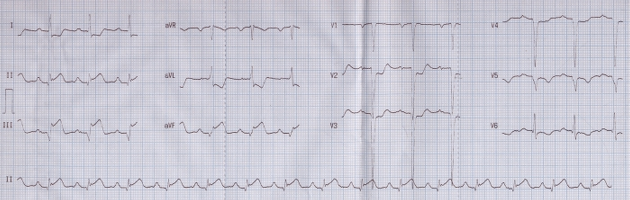
Q waves and ST segment elevation in inferior leads II, III and aVF, with reciprocal ST segment depression in leads I, aVL, V2-V4 is suggestive of acute inferior wall myocardial infarction. Negative P waves in lead V1 is indicative of left atrial overload. QS complexes in leads V1 to V5 is in favor of old anterior wall myocardial infarction.
The upsloping ST elevation with upright T waves in inferior leads are a feature of hyperacute inferior wall infarction. This case will benefit from thrombolytic therapy or primary angioplasty. Coronary angiography is likely to reveal a previous left anterior descending coronary artery lesion and a fresh occlusion of right coronary artery. ST elevation in lead III is more than that in lead II, which also favors right coronary occlusion, rather than left circumflex occlusion, in which you expect the ST elevation in lead II to be more than that in lead III.
Related Posts

About The Author
Johnson Francis
Former Professor of Cardiology, Calicut Govt. Medical Kozhikode, Kerala, India. Editor-in-Chief, BMH Medical Journal