ECG Quiz with discussion – Pacing
ECG Quiz with discussion – Pacing
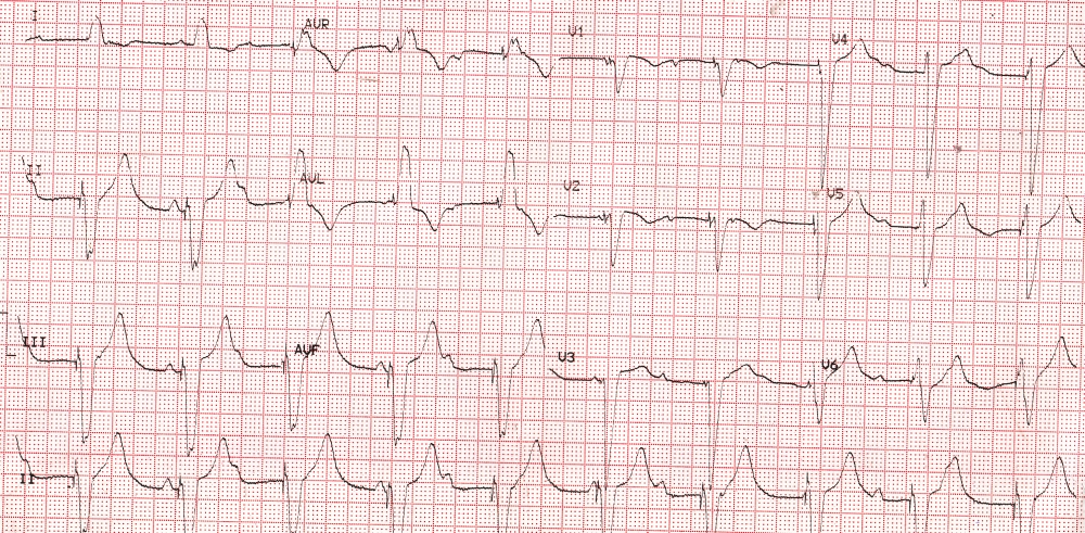
What are the important findings and diagnosis?
ECG shows a regular wide QRS rhythm at a rate of 60/minute. Each QRS complex is preceded by a narrow spike indicating ventricular paced rhythm. Dissociated P waves are seen suggesting that it is a single chamber ventricular pacing. Left bundle branch block pattern in I and aVL would mean right ventricular pacing. Inferior leads show negative QRS complexes indicating spread of activity from below upwards, suggesting right ventricular apical pacing. Right ventricular pacing causes left ventricular dyssynchrony and can cause left ventricular systolic dysfunction and heart failure in the long run. Lack of AV synchrony in single chamber pacing will manifest as intermittent cannon waves in the jugular venous pulse.
Related Posts

About The Author
Johnson Francis
Former Professor of Cardiology, Calicut Govt. Medical Kozhikode, Kerala, India. Editor-in-Chief, BMH Medical Journal