Echocardiographic profile in aortic stenosis – Echocardiogram video
Echocardiographic profile in aortic stenosis – Echocardiogram video
Echocardiogram with narration:
Echocardiogram without narration:
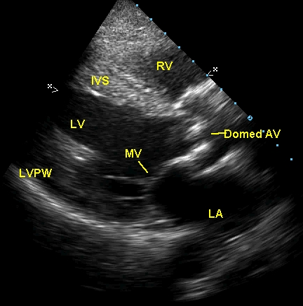
Echocardiogram in parasternal long axis view showing calcific aortic valve which domes in systole with restricted opening. The aortic orifice is quite narrow. Normally valve leaflets will be almost invisible in systole as they will get apposed to the walls of the aortic sinuses of Valsalva. Doming occurs due to commissural fusion, which restricts systolic opening movements. LA: left atrium; Domed AV: domed aortic valve; MV: mitral valve (in closed position); RV: right ventricle; IVS: interventricular septum; LV: left ventricle; LVPW: left ventricular posterior wall. Concentric left ventricular hypertrophy is associated. The rounded echolucency behind the left atrium is the cross section of the descending aorta.
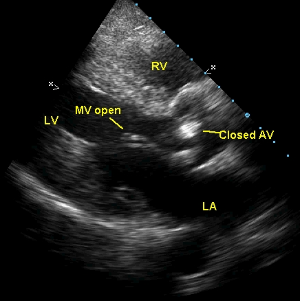
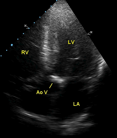
Diastolic frame in aortic stenosis from parasternal long axis view showing aortic valve in closed position (closed AV) and mitral valve (MV open) in open position. The marker is indicating the anterior mitral leaflet. Dense calcification of aortic valve is evident.
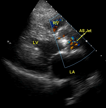
Colour flow mapping (CFM, color Doppler imaging) from parasternal long axis view in aortic stenosis shows the domed aortic valve with narrow aortic orifice and a mosaic (multicolored) jet (AS Jet) beyond the valve, indicating turbulent flow due to the stenosis. Note that the left ventricular cavity (LV) is quite small in systole due to the hypertrophy.
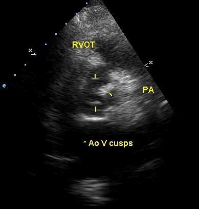
Parasternal short axis view in calcific aortic stenosis, showing the three leaflets in open position (systolic frame). The markers show the leaflets (Ao V cusps). The commissure between the left and right coronary cusps appear fused and thickened. RVOT: right ventricular outflow tract; PA: pulmonary artery.
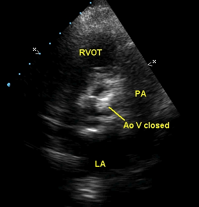
Parasternal short axis view at the level of the aortic valve showing the aortic valve in closed position (Ao V closed) with significant thickening and calcification. The valve appears trileaflet rather than bicuspid.
Apical five chamber (apical 5C) view in echocardiography showing the thickened aortic valve (Ao V). This is the usual view used for estimating the trans valvar gradients as the Doppler beam is parallel to the jet in this view. Another view used for estimating the gradient is the suprasternal view, especially when associated mitral regurgitation gets picked up by the Doppler beam giving a falsely high gradient from the apical 5C view.
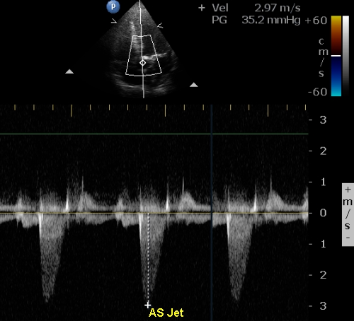
Color flow guided evaluation of aortic stenosis jet from apical five chamber view with continuous wave Doppler interrogation. The upper panel shows the color Doppler image and the Doppler line. The color bar at the top right shows the Nyquist limit of the color flow mapping system (60 cm/s in this case). The lower panel shows the tongue shaped envelope of valvar aortic stenosis (contrast from the sickle shaped envelope of signal in case of left ventricular outflow obstruction). The peak velocity in this case is 2.97 m/s which corresponds to a gradient of 35.2 mm Hg. Since the aortic valve shows significant calcification, it is possible that acoustic shadowing is preventing the localisation of the jet region with maximum velocity. Hence another evaluation of the gradient from the suprasternal view is needed.
Related Posts


About The Author
Johnson Francis
Former Professor of Cardiology, Calicut Govt. Medical Kozhikode, Kerala, India. Editor-in-Chief, BMH Medical Journal