Echocardiographic profile in bioprosthetic mitral valve
Echocardiographic profile in bioprosthetic mitral valve

Echocardiogram of bioprosthetic mitral valve in parasternal long axis viewEchocardiogram of bioprosthetic mitral valve in parasternal long axis view showing the valve in open position. RV: right ventricle; Ao: aorta; LV: left ventricle; LA: left ventricle. Bioprosthetic valves can be with or without stents. This image is from a stented bioprosthetic valve. The porcine valve is fixed in glutaraldehyde. The low profile stent is made up of an acetyl homopolymer. Stentless valves have larger orifice than stented valves.
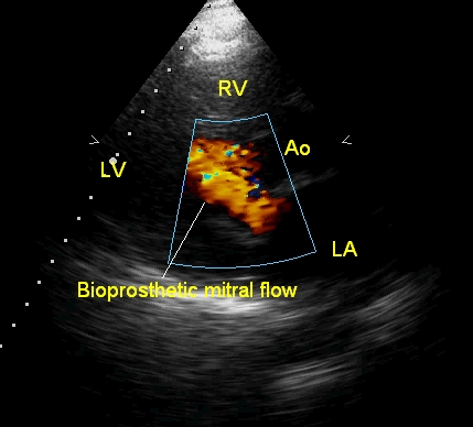
Fairly laminar across a bioprosthetic mitral valve in parasternal long axis view – most of the flow is in single colour though some variance is seen. Small areas of turbulence are seen as mosaic (multi-coloured) regions. Left atrial diameter in this view is slightly more than that of the aorta, indicating residual left atrial dilatation from the mitral valve disease.
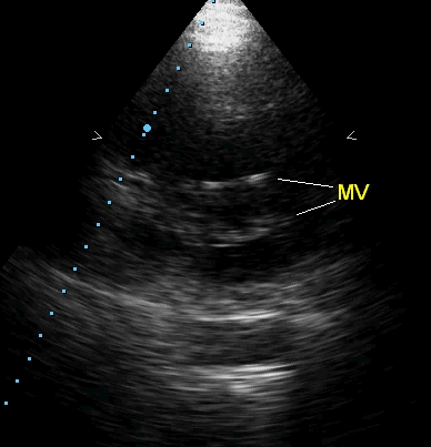
Short axis view of bioprosthetic mitral valve, apparently simulating a mild rheumatic mitral stenosis, due to the echodensity of the valve stent. But there is a good central orifice.
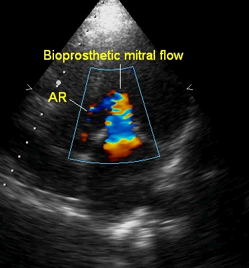
Bioprosthetic mitral flow as seen from apical four chamber view: The flow is slightly turbulent in this view, possibly due to the different settings of Nyquist limit due to changes in depth of imaging compared to the parasternal long axis view. A trivial aortic regurgitation (AR) jet is also seen.
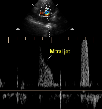
Mitral flow on pulsed Doppler interrogation, indicating that the velocities and gradients are not high across the bioprosthetic valve which is functioning well. Normal gradients across a prosthetic valve are higher than that across a normal valve, but varies depending on the type of the prosthesis and the location. Prosthetic valves at the aortic position can have a higher gradient than at the mitral position, basically because of the smaller orifice and the higher force of ventricular ejection.
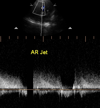
Aortic regurgitation (AR) jet on continuous wave Doppler: The jet is incompletely seen as the volume of regurgitation is only trivial. Normal aortic forward flow is seen as a small spectrum below the baseline. Continuous wave interrogation is used because the velocity of an aortic regurgitation jet is much above the Nyquist limit of the pulsed Doppler system. Continuous wave Doppler can evaluate high velocity jets, but cannot localise the exact site of the jet along the axis.
Related Posts


About The Author
Johnson Francis
Former Professor of Cardiology, Calicut Govt. Medical Kozhikode, Kerala, India. Editor-in-Chief, BMH Medical Journal