EP tracing during ventricular pacing
EP tracing during ventricular pacing
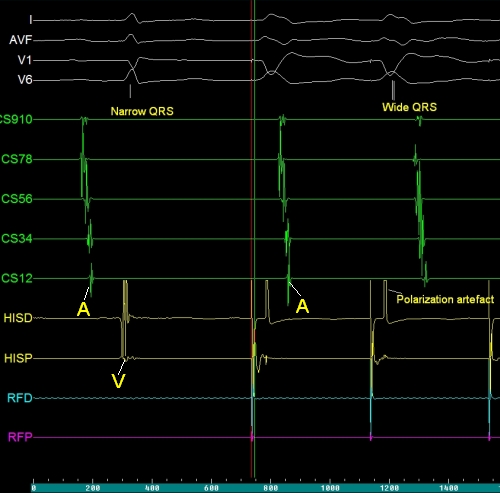
EP tracing during ventricular pacing showing atrial and ventricular activations. The CS 910 electrode is in the proximal coronary sinus while the CS12 electrode is in the distal coronary sinus. ‘A’ represents atrial activation and ‘V’ represents ventricular activation. The pacing stimulus is delivered from the HISD electrode as evidenced by the polarization artefact seen in this lead. But the electrode does not seem to be in the region of the His bundle as neither the His bundle activity nor the atrial activity is seen as is usual. It seems that the lead has been pushed into the ventricle during the EP study for ventricular pacing. This is sometimes done to minimise the total number of electrodes being used during an EP study. The RFD and RFP electrodes do not seem to be connected to the recorder as these electrodes are not recording any electrical activity of the heart. The initial QRS is narrow while the subsequent two QRS complexes preceded by pacing spikes are wide. The first one is a sinus beat conducted to the ventricles while the latter two are paced beats. The atrial activity picked up by the coronary sinus leads show almost the same sequence of activation as the sinus beat, though it is retrogade activation. But the retrograde interval is longer for the second paced beat compared to the first paced beat.
Related Posts

About The Author
Johnson Francis
Former Professor of Cardiology, Calicut Govt. Medical Kozhikode, Kerala, India. Editor-in-Chief, BMH Medical Journal