Left bundle branch block (LBBB) with 2:1 AV block
Left bundle branch block (LBBB) with 2:1 AV block
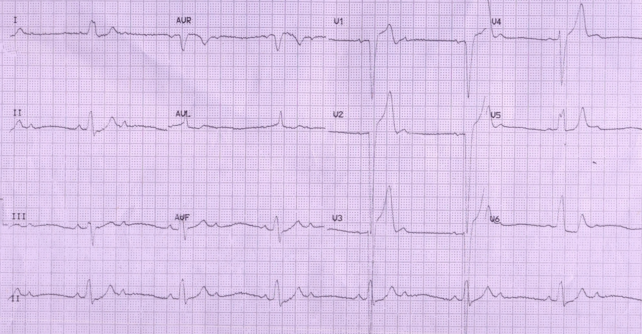
Left bundle branch block (LBBB) is manifested as a wide QRS with M shaped pattern in lateral leads and wide QS complexes in anterior leads. The initial q waves are absent in lateral leads due to the loss of initial left to right activation of the septum due to left bundle branch block. For the same reason, the initial r wave is missing in anterior leads. There are two P waves for every QRS complex, with atrial rate double that of ventricular rate, denoting 2:1 atrioventricular (AV) conduction (2:1 AV block). Together with LBBB, this constitutes one form of trifascicular block.
The presence of LBBB indicates that the block is infra Hisian. In fact a subsequent ECG showed complete heart block, making the indication for permanent pacing more urgent. Usually the ST segment and T waves are discordant in LBBB (opposite the predominant QRS deflection). Here the T waves in lateral leads are upright (concordant), indicating a primary T wave abnormality). Associated coronary artery disease or myocardial disease has to be thought of in this setting. Tall T waves are noted in lead V2 – V3. QS complexes are noted in V1 and V2. rS pattern is seen in V3. There is upsloping ST depression in leads V1-V4. ST is isoelectric in V5 and there is ST depression in V6.
Related Posts


About The Author
Johnson Francis
Former Professor of Cardiology, Calicut Govt. Medical Kozhikode, Kerala, India. Editor-in-Chief, BMH Medical Journal