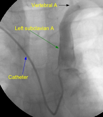
Left subclavian angiogram to visualise LIMA
Left subclavian angiogram to visualise LIMA

Left subclavian angiogram
Left subclavian angiogram by trans-radial route using tiger catheter. Left subclavian angiogram is usually taken towards the end of coronary angiography procedure when multivessel disease or left main coronary artery disease requiring coronary artery bypass grafting is documented. This is in order to visualize the left internal mammary artery to be used for bypass grafting. This is an early frame so that the LIMA is not clearly seen. Vertebral artery is seen faintly, arising from the left subclavian, at the upper part of the image.
In femoral approach, left subclavian angiogram is usually taken with the Judkin’s right coronary catheter which can be pushed further from the upper descending aorta to cannulate the left subclavian artery. This is usually done while withdrawing the JR catheter after right coronary angiogram. The convenience is that we don’t have to use another catheter for cannulating the left subclavian artery. Opacification of LIMA can be improved by compressing the left brachial artery externally to reduce runoff into the left upper limb during left subclavian injection.
It is also possible to inject directly into LIMA by pushing the JR catheter further into the left subclavian and gradually pulling back. But direct cannulation of LIMA is seldom needed as non selective left subclavian injection gives reasonable opacification of the LIMA. Super-selective cannulation of LIMA may be needed in rare cases due to poor opacification with left subclavian injection. Care has to be taken to avoid damage to the LIMA ostium during superselective injections. Special catheters for cannulating the LIMA are also available. The person may experience discomfort in the chest during a LIMA injection, especially with the older variety of ionic contrast agents.

