Left ventricular angiogram in right anterior oblique view
Left ventricular angiogram in right anterior oblique view
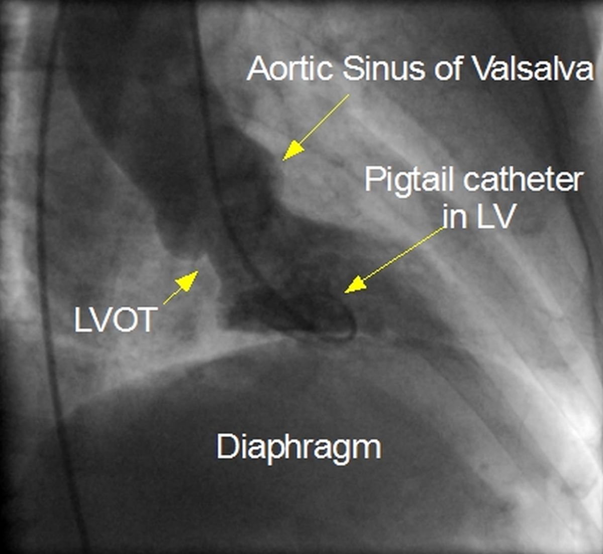
LVOT: Left ventricular outflow tract; LV: Left ventricle; RAO: Right anterior oblique
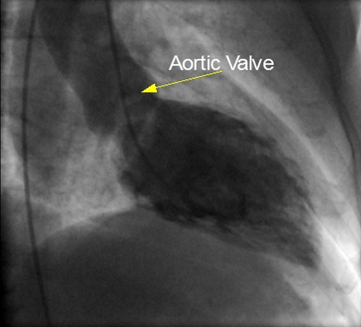
Left ventricular angiogram, systolic frame in RAO view
Pigtail catheter is seen in the left ventricular cavity. Pigtail catheter is used for pressure injection because it is less likely to produce ventricular ectopy and contrast staining of the myocardium. Contrast staining if severe can lead to refractory ventricular arrhythmias rarely. As there are multiple holes in different directions, the recoil force on the pigtail catheter is cancelled out and the chance of a single jet producing myocardial staining is reduced. The pigtail should be in the cavity and not in the inflow or outflow region. If it is in the inflow region, it can produce mitral regurgitation. A ventricular ectopy induced by the catheter can also produce a whiff of mitral regurgitation. Left ventricular angiogram (left ventriculogram) in RAO view is usually taken to check the regional wall motion, left ventricular function and mitral regurgitation if any. The catheter is visible in the ascending (contrast filled) and descending (partly contrast filled) aorta. This means that the pigtail catheter was introduced through the transfemoral route. Contrast filled bulges in the root of aorta are the aortic sinuses of Valsalva. Silhouette of the diaphragm is seen just below the cardiac shadow.
Left ventricular angiogram, diastolic frame in RAO view
The diastolic frame of the left ventricular angiogram shows trabeculae carneae as negative shadows. The aortic valve in the closed position is seen as a thin negative shadow at the base of the sinus of Valsalva. Negative shadow of mitral diastolic flow can be seen at the posterobasal region. Thickness of the ventricular wall can be assessed by noting the distance between the contrast filled region (cavity) and the cardiac silhouette, especially in the anterolateral region.
Related Posts

About The Author
Johnson Francis
Former Professor of Cardiology, Calicut Govt. Medical Kozhikode, Kerala, India. Editor-in-Chief, BMH Medical Journal