Mitral E/E’ for assessment of left ventricular diastolic function
Mitral E/E’ for assessment of left ventricular diastolic function
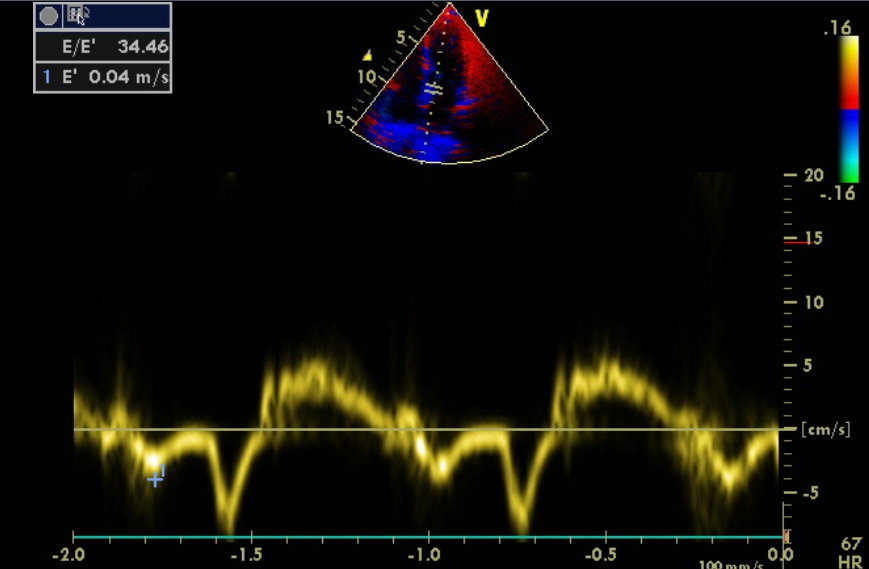
E/E’ measured using a combination of mitral flow Doppler and tissue Doppler of mitral annulus is an important measure of left ventricular diastolic function. Assessment of diastolic function of the left ventricle is assuming more importance after the recognition of diastolic heart failure which is otherwise known as heart failure with preserved ejection fraction (HFpEF). As the population ages, frequency of HFpEF is on the rise. The image illustrates measurement of E’ by tissue Doppler. Previously measured E is used to calculate the E/E’ ratio here. Tissue Doppler measurement in the image is from the medial mitral annulus region.
Conventionally reversal of E/A (early diastolic and atrial systolic mitral flow velocities) is taken as indicating left ventricular diastolic dysfunction. In normal left ventricular flow pattern, E wave is taller than A wave. With diastolic dysfunction, early diastolic flow decreases and the late diastolic flow gets augmented due to more forceful atrial contraction.
Mitral flow varies both with left atrial pressure and the left ventricular relaxation time constant (tau). E’ measured by tissue Doppler is the movement of mitral annulus during early diastole and is inversely proportional to tau. Hence E/E’ will be directly proportional to left atrial pressure. Hence E/E’ will be a good indicator of left ventricular filling pressure [1].
E/E’ ratio above 15 is taken as an indicator of left ventricular diastolic dysfunction. Here it is well above that (34.46). A value below 8 can be considered as normal value intermediate values will be in the grey zone.
A study in 15 ambulatory chronic heart failure patients with an implanted device to measure left atrial pressure directly, E/E’ had the greatest accuracy for detection of left atrial pressures of 15 mm Hg or more [2]. Mitral E velocity and E/A ratio had lesser accuracy.
E/E’ is useful in the setting of atrial fibrillation where E/A ratio is not applicable for assessment of left ventricular diastolic function.
Downside of E/E’ in assessing left ventricular diastolic function
It has been mentioned that E/E’ may not be an ideal measurement to indicate left ventricular filling pressures in decompensated patients with advanced systolic heart failure. Difficulty has been noted in those with large left ventricular volumes and impaired cardiac indices as well as in the presence of cardiac resynchronization therapy [3].
References
- Park JH, Marwick TH. Use and Limitations of E/e’ to Assess Left Ventricular Filling Pressure by Echocardiography. J Cardiovasc Ultrasound. 2011 Dec;19(4):169-73.
- Ritzema JL, Richards AM, Crozier IG, Frampton CF, Melton IC, Doughty RN, Stewart JT, Eigler N, Whiting J, Abraham WT, Troughton RW. Serial Doppler echocardiography and tissue Doppler imaging in the detection of elevated directly measured left atrial pressure in ambulant subjects with chronic heart failure. JACC Cardiovasc Imaging. 2011 Sep;4(9):927.
- Mullens W, Borowski AG, Curtin RJ, Thomas JD, Tang WH. Tissue Doppler imaging in the estimation of intracardiac filling pressure in decompensated patients with advanced systolic heart failure. Circulation. 2009 Jan 6;119(1):62-70. (Free full text at Pubmed Central).
About The Author
Johnson Francis
Former Professor of Cardiology, Calicut Govt. Medical Kozhikode, Kerala, India. Editor-in-Chief, BMH Medical Journal