ECG of pacing through lateral cardiac vein
ECG of pacing through lateral cardiac vein
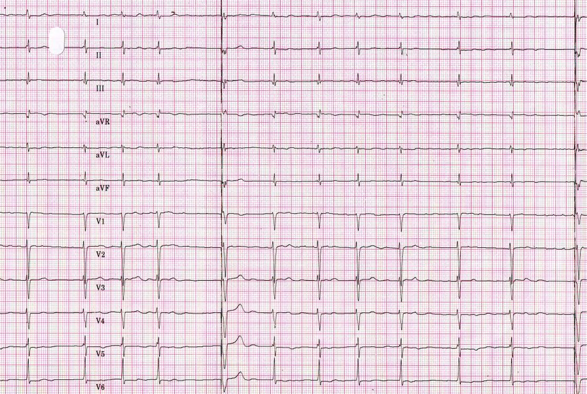
Pacing through lateral cardiac vein was done because of prior triple valve replacement, mitral, tricuspid and aortic, which precluded transvenous right ventricular pacing. The basic rhythm is atrial fibrillation and the fifth and last beats are paced. The tall spikes before the paced QRS complexes indicate the possibility of unipolar pacing which causes tall pacing artefacts.
QS complexes in leads V4-V6 indicate an activation proceeding medially from the lateral wall of the left ventricle. This is quite different from the activation seen with the basic rhythm.
Related Posts

About The Author
Johnson Francis
Former Professor of Cardiology, Calicut Govt. Medical Kozhikode, Kerala, India. Editor-in-Chief, BMH Medical Journal