Latest
ECG / Electrophysiology
The two common methods of mapping cardiac arrhythmias are activation mapping and pace mapping.
Read More
Angiography and Interventions
Catheter induced ectopy is due to irritation of the local endocardium and will show all the features of a target ventricular ectopic for ablation.
Read More
ECG / Electrophysiology
ST elevation during treadmill test documented by serial ECGs through recovery.
Read More
ECG / Electrophysiology
ECG showing Sinus bradycardia with first degree AV block.
Read More
ECG / Electrophysiology
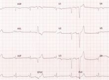
ECG showing Ventricular ectopic beats.
Read More
ECG / Electrophysiology
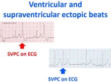
Ventricular ectopic beats are recognized as wide bizarre QRS complexes which occur prematurely and are not usually preceded by a P wave.
Read More
ECG / Electrophysiology
Ventricular fibrillation is recognized on the electrocardiographic monitor as a highly disorganized rhythm with no definite P waves or QRS complexes.
Read More
ECG / Electrophysiology
Three or more ventricular complexes (wide QRS) occurring at a rate more than 100 per minute is taken as ventricular tachycardia (VT).
Read More
ECG / Electrophysiology
Accelerated idioventricular rhythm (AIVR) is a classical reperfusion arrhythmia which occurs during thrombolysis of acute myocardial infarction.
Read More
ECG / Electrophysiology
Three or more different morphologies of P waves, with a rate more than 100 per minute defines multifocal atrial tachycardia (MAT) or chaotic atrial rhythm.
Read More
Posts navigation