Proximal left coronary artery on echocardiogram
Proximal left coronary artery on echocardiogram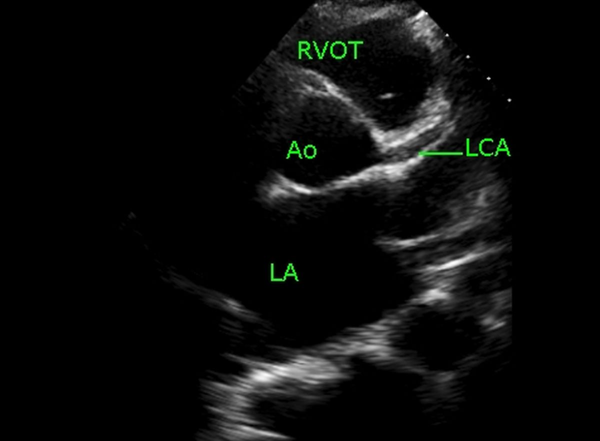
Proximal portion of major coronary arteries can often be seen on echocardiography, while the distal regions can seldom be imaged. Coronary arteries are better seen if they are dilated, especially in those with aneurysms in Kawasaki disease. In this case proximal left coronary artery is mildly dilated, possibly because of underlying valvular lesions with left ventricular dilatation and hypertrophy.
Coronary arteries are relatively easier to image in children, while in adults echo window is often not good enough for imaging coronary arteries by echocardiography. In some patients with renal failure, the images are good enough so that left anterior descending coronary artery can be imaged to a long extent. Probably the waterlogged thorax permits better transmission of ultrasound in these cases.
A detailed guide on how to image all coronary arteries by echocardiography has been published by Marek Krzanowski and colleagues [1]. The article contains numerous echocardiographic images with Colour Doppler and Doppler in various views. In addition multiple correlative coronary angiograms are also provided. Anatomical pictures have also been given.
Precautions like how to avoid mistaking extra cardiac vessels and a cardiac vein while imaging the mid portion of left anterior descending coronary artery has been mentioned. Spectral Doppler will show typical predominantly systolic flow in extra cardiac arteries, which is seen in other peripheral arteries. Veins will show low velocity continuous flow which will stop or decrease significantly with Valsalva maneuver. Coronary arterial flow is predominantly diastolic.
Reference
- Krzanowski M, Bodzoń W, Dimitrow PP. Imaging of all three coronary arteries by transthoracic echocardiography. An illustrated guide. Cardiovasc Ultrasound. 2003 Nov 17;1:16.
Related Posts


About The Author
Johnson Francis
Former Professor of Cardiology, Calicut Govt. Medical Kozhikode, Kerala, India. Editor-in-Chief, BMH Medical Journal