RHD MR and AR on colour Doppler echo
RHD MR and AR on colour Doppler echo
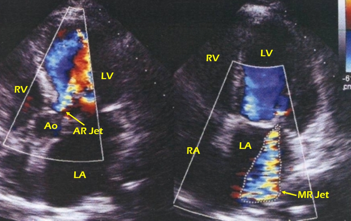
Colour Doppler echocardiogram in rheumatic heart disease (RHD), mitral regurgitation (MR) and aortic regurgitation. Left frame shows aortic regurgitation jet from aorta to left ventricle. It appears to be at least moderate severity. LA: Left atrium; RA: Right atrium; LV: Left ventricle; RV: Right ventricle; Ao: Aorta; AR Jet: Aortic regurgitation jet; MR Jet: Mitral regurgitation jet.
The mosaic coloured MR jet is outlined in the left atrium (right frame). The left frame shows the apical five chamber view in diastole, while the right frame shows the apical four chamber view in systole. The apical five chamber view shows an aortic regurgitation jet which merges with the mitral jet in the left ventricle. Mitral leaflets appear thickened, though the finer morphological details of the mitral and aortic valves are not clear in these colour flow mapping images. MR severity can be assessed by comparing the MR jet area with that of the left atrium as well as by observing the extent of penetration of the jet into the left atrium. The larger the comparative jet area and more distant the penetration of the jet, higher the severity of the mitral regurgitation. A disadvantage for comparing the area of the jet with that of the left atrium is in the presence of a grossly dilated left atrium. Similarly, the depth of jet penetration may vary depending on the eccentricity of the jet. This is also applicable to jet area, with eccentric jet appearing smaller in area.
Related Posts


About The Author
Johnson Francis
Former Professor of Cardiology, Calicut Govt. Medical Kozhikode, Kerala, India. Editor-in-Chief, BMH Medical Journal