Flail posterior mitral leaflet with mitral regurgitation
Flail posterior mitral leaflet with mitral regurgitation
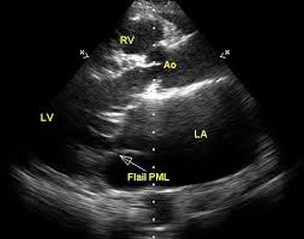
Two dimensional (2D) echocardiographic image showing right ventricle (RV), aorta (A0), left atrium (LA), left ventricle (LV) and the flail posterior mitral leaflet (PML) which points back into the left atrium during systole. The gap between the posterior and anterior leaflets is evident and leads to severe mitral regurgitation (below).
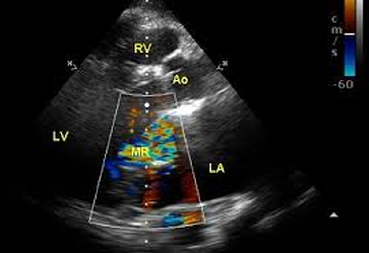
Colour flow mapping (colour Doppler) superimposed on a 2D echocardiogram showing the multi coloured (mosaic) jet of mitral regurgitation (MR) into the left atrium, behind the anterior mitral leaflet and anterior to the flail PML. Since the jet is directed anteriorly, the systolic murmur of mitral regurgitation in this case will be conducted to the base of the heart (medially and upwards from the apex) rather than to the axilla as in classical rheumatic mitral regurgitation. Even though the frame shown here demonstrates only a small area of the jet, multiple projections confirmed that it is severe mitral regurgitation. Flail mitral leaflet occurs due to rupture of the chordae tendineae, which could be spontaneous or secondary to infective endocarditis or other inflammatory pathology. Chordal rupture can also occur with myocardial infarction. Lijun Cui, Ya Suo, Liju Han, Zhao Li, Xianghong Ma, Changyu Zhou and Guangping Li reported a case of rupture of mitral chordae tendineae in acute inferior wall myocardial infarction [1].
Reference
- Lijun Cui, Ya Suo, Liju Han, Zhao Li, Xianghong Ma, Changyu Zhou, Guangping Li. Ruptured mitral chordae tendinae induced by acute inferior myocardial infarction. Int J Cardiol. 2015 Feb 15;181:216-7.
About The Author
Johnson Francis
Former Professor of Cardiology, Calicut Govt. Medical Kozhikode, Kerala, India. Editor-in-Chief, BMH Medical Journal