RHD MS PSAX view
RHD MS PSAX view
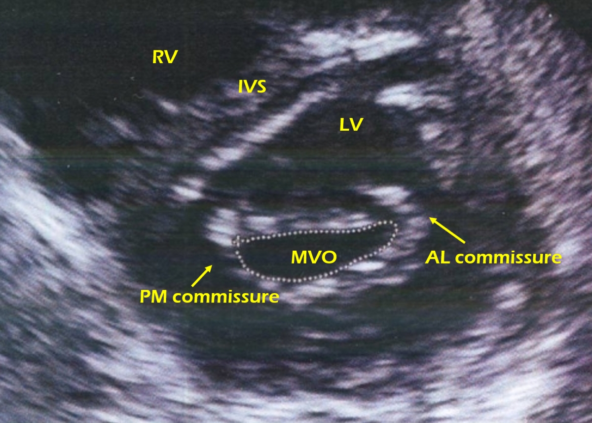
RHD MS PSAX view: Parasternal short axis view showing mitral valve in cross section (dotted outline). The valve leaflets are thickened and the commissures are fused. The cut is slightly oblique as a good cut should appear circular. It is often difficult to get a good circular outline of the mitral valve to varying anatomical features of the chambers and cardiac position. Ideally the smallest full circle should be taken to planimeter the valve area. If it is not a full circle, the subvalvar pathology may be measured as the valve orifice. If the smallest full circle is not taken, it will be the valve proximal to the severest stenosis (valve belly). The mitral valve cross section in mitral stenosis has a fish mouth appearance. Here the commissures are fused, though there is some echo drop outs, which is there in the belly of the valve leaflets as well. The left ventricular cavity surrounds the mitral orifice, which in turn is surrounded by the left ventricular wall. Interventricular septum with the right ventricle beyond is seen towards the upper left corner. IVS: Interventricular septum; RV: Right ventricle; LV: Left ventricle; AL commissure: Anterolateral commissure; PM: Posteromedial commissure; MVO: Mitral valve orifice. RHD: Rheumatic heart disease; MS: Mitral stenosis; PSAX: Parasternal short axis view.
Related Posts

About The Author
Johnson Francis
Former Professor of Cardiology, Calicut Govt. Medical Kozhikode, Kerala, India. Editor-in-Chief, BMH Medical Journal