Rheumatic mitral stenosis – echocardiogram in PLAX view
Rheumatic mitral stenosis – echocardiogram in PLAX view
Rheumatic mitral stenosis – echocardiogram in PLAX viewMitral valve is the most commonly involved valve in rheumatic heart disease. According to Paul Wood, the involvement of the valves in rheumatic heart disease is depending on the hemodynamic load on the valve. The mitral valve faces the maximum load as it withstands the contractile force of the left ventricle during systole. The next highest load is on the aortic valve, the load on which is the aortic diastolic pressure.
Rheumatic mitral valve involvement is characterised by commissural fusion. This causes the valve leaflets to move together in the same direction during diastole. Normally the anterior mitral leaflet (AML) moves anteriorly during the opening motion in diastole and the posterior leaflet moves posteriorly. Due to commissural fusion the posterior leaflet moves anteriorly along with the anterior mitral leaflet in diastole. This is a paradoxical movement of the posterior mitral leaflet (PML). The movement of the anterior mitral leaflet is also restricted, producing a doming or hockey stick like appearance in diastole. The diastolic separation between the leaflets is also reduced, obstructing the forward flow across the valve. In cross sectional imaging the mitral valve has a fish mouth appearance, with thickening of both leaflets and fusion of anterolateral and posteromedial commissures.
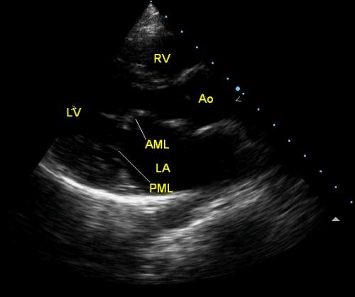
The image above illustrates all the feature in the parasternal long axis view (PLAX) like doming of AML, anterior displacement of the PML and reduced leaflet separation in diastole. The dilated left atrium (LA) secondary to the mitral valve obstruction is also evident. The circular echolucency behind the left atrium is the cross section of the descending aorta. In live imaging it will seen as pulsatile, increasing dimensions in systole and reducing in diastole. Ascending aorta (Ao) is seen anterior to the left atrium and is of normal size. Aortic valve is in a closed position and is seen only faintly as it is not thickened. Left ventricular cavity is seen distal to the mitral valve (LV) and the right ventricle (RV) is seen above, with the interventricular septum in between (not marked). The apex of the triangular image is the position of the transducer, which has been kept in the left parasternal region, on the anterior chest wall, just anterior to the right ventricle.
Related Posts


About The Author
Johnson Francis
Former Professor of Cardiology, Calicut Govt. Medical Kozhikode, Kerala, India. Editor-in-Chief, BMH Medical Journal